A description of the muscles of the human body, as they appear on dissection : with the synonyma of Cowper, Winslow, Douglas, Albinus, and Innes, and the new nomenclature of Dumas, professor of anatomy at Montpellier : with prints and maps, showing the insertions of muscles / by Joseph Constantine Carpue.
- Joseph Constantine Carpue
- Date:
- 1801
Licence: Public Domain Mark
Credit: A description of the muscles of the human body, as they appear on dissection : with the synonyma of Cowper, Winslow, Douglas, Albinus, and Innes, and the new nomenclature of Dumas, professor of anatomy at Montpellier : with prints and maps, showing the insertions of muscles / by Joseph Constantine Carpue. Source: Wellcome Collection.
Provider: This material has been provided by The Royal College of Surgeons of England. The original may be consulted at The Royal College of Surgeons of England.
65/90 (page 39)
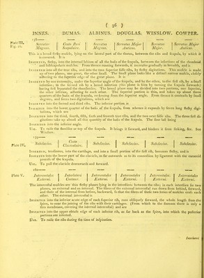
![INNES. 167 Rectus Capitis Interims Major. COWPER. Rectus Interims Major. Inserted into the anterior points of the tranfverfe procefles of the third, fourth, fifth, and fixth cervical ver- tebrae, and is Inserted into the cuneiform procefs of the os occipitis, before the condyloid procefs. Use. To bend the head forward. 168 DUMAS. ALBINUS. DOUGLAS. WINSLOW. Grand Rectus Capitis Reetus Rectus Capitis 1 rach’elo Internus Internus Anticus Basilaire. Major. Major Longus. Rectus Capitis Internus Minor. Petit Rectus Capitis. Rectus Rectus Trachelo Internus Internus Anticus Basilaire. Minor. Minor. Brevis. Rectus Interims Minor. Inserted, fiethy, into the fore part of the body of the firft cervical vertebra, and is Inserted into the root of condyloid procefs of the os occipitis, under and more outwards than the laft mufcle. Use. To bend the head forwards. Rectus Capitis Lateralis. Tracheli Rectus Reetus Transversal is i; Altoido Capitis Lateralis Anticus 1 Basilaire. Lateralis. Fallop. Primus. Rectus Lateralis. Inserted, flefliy, into the tranfverfe procefs of the firft cervical vertebra, near its extremity, and is Inserted into the os occipitis, oppofite to the foramen ftylo maftoideum. Use. To bend the head a little to one fide. 170 Psoas Magnus.* Pre Psoas, sive Lumbo j Psoas Magnus. Psoas Magnus. Lumbaris Troehantin. | Internus. Psoas Magnus. This is a long thick mufcle. The upper infertions are in the abdomen, on the lumbar region, the lower infec- tion is in the thigh. It is -p 0 Inserted, flefliy, into the fides and tranfverfe procefles of all the lumbar vertebra?, by diftimft flips From thele mfertions it runs down laterally over the ilium, on the fide of the iliacus internus, to which it is con- nected ; pafTes under Poupart’s ligament, covers the fore fide of the head of the os femoris, and is Inserted, tendinous, into the fore part of the little trochanter. Use. Bends the thigh forwards, or when the lower extremity is fixed, aflifts in bending the body. Iliacus * ^e-V'?US t0 H16 dercr,'Pt;° °,f, mufclcs of the thigh, it is neceflary to underftand the ftru&ure and infertions of the fafri- tern a 1 I^ ef* a11.lhe mufc,eS of the ,hi8h alld “ » tufcular ligament, made up of two planed• the« ternal more or lefs longitudinal, the internal more or lefs tranfverfe It is fhengthened in iome placesbv a iimnWr^f of.u’ £k which augment us thicknefs, and foim particular expanfions. it is inferred above into the edge ofPthe crifta olEs ilium into the 1 tuberefity to the anterior fuperior fpinous procefs, into Paupart’s ligament, and to the aponeurofis h t nhl n ” k ■2 it rims up by a thin lamina, is infected into the lateral inferior part of the os fecrum and ^ ^uons it advance ^ the glutai a9d thigh, between the membrana adipofa and mufcle. I the thi knee. It is very thin on the patella. It is continued over the external anterior part of the tibia is inferred ism the InU n TV part 0f the fibula; U fendS 0ff «-“*«“ions, r.i^^bman ^un1^‘i tt^ muf S’mduiUvt U V a maTfr’ ,as t0 f0rm vagmf' ]tu ftron8er on the auterior and7 outer parts of the thigh, grow- n . thinner on the inner and hack parts. It is inferted into the iinea afpera, between the vaftus externus and biceS-° it chieflrmaPderuJofa'trSfe£ ' h * °D °f ^ thi«h* Th°Ugh ,hefc vaSin* thin, they are ftrong/being t It is fometimes inferted into the body of the firft dorfal vertebra. Plate III. Fig. 12, Plate III. Fig. iy Plate III. Fig 13.](https://iiif.wellcomecollection.org/image/b22415592_0067.jp2/full/800%2C/0/default.jpg)