A text-book of the diseases of the ear for students and practitioners / by Adam Politzer ; tr. at the personal request of the author and ed. by Milton J. Ballin and Clarence L. Heller.
- Ádám Politzer
- Date:
- 1903
Licence: Public Domain Mark
Credit: A text-book of the diseases of the ear for students and practitioners / by Adam Politzer ; tr. at the personal request of the author and ed. by Milton J. Ballin and Clarence L. Heller. Source: Wellcome Collection.
Provider: This material has been provided by the Francis A. Countway Library of Medicine, through the Medical Heritage Library. The original may be consulted at the Francis A. Countway Library of Medicine, Harvard Medical School.
34/912 page 12
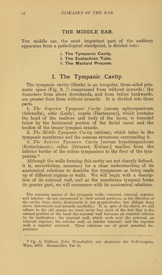
![THE MIDDLE EAR. The middle ear, the most important part of the auditory apparatus from a pathological standpoint, is divided into: 1. The Tympanic Cavity. 2. The Eustachian Tube. 3. The Mastoid Process. I. The Tympanic Cavity. The tympanic cavity (Henle) is an irregular, three-sided pris- matic space (Fig. 9, /) compressed from without inwards; the diameters from above downwards, and from before backwards, are greater than from without inwards. It is divided into three parts. 1. The Superior Tympanic Cavity [cavum epitympanicum (Schwalbe), attic (Leidy), cupola (Hartmann)], which contains the head of the malleus and body of the incus, is bounded below by the horizontal portion of the facial canal and the tendon of the tensor tympani muscle. 2. The Middle Tympanic Cavity (atrium), which takes in the tympanic membrane and the osseous structures surrounding it. 3. The Inferior Tympanic Cavity [cavum hypotympanicum (Kretschmann), cellar (Grunert, Korner)] reaches from the inferior border of the sulcus tympanicus to the floor of the tym- panum.* Although the walls forming this cavity are not sharply denned, it is, nevertheless, necessary for a clear understanding of its anatomical relations to describe the tympanum as being made up of different regions or walls. We will begin with a descrip- tion of its external wall, and as the membrana tympani forms its greater part, we will commence with its anatomical relations. The common names of the tympanic walls—external, internal, superior and inferior—do not correspond to their actual positions, as the direction of the cavity from above downwards is not perpendicular, but oblique from above downwards and inwards (medially). If, in spite of the above, we still adhere to the old names, we must never lose sight of the fact that in the normal position of the head the external wall becomes an external inferior by its inclination; the internal wall, which roofs over the external, an internal superior; the inferior wall, an inferior internal; and the superior wall, a superior external. These relations are of great practical im- portance. * Cp. A. Politzer, Zelin Wandtafeln zur Anatomie des Oehororgans. Wien, 1873. Braumiiller, Taf. iii.](https://iiif.wellcomecollection.org/image/b21169494_0034.jp2/full/800%2C/0/default.jpg)


 No text description is available for this image
 No text description is available for this image
 No text description is available for this image