Licence: Public Domain Mark
Credit: Human anatomy (Volume 1). Source: Wellcome Collection.
Provider: This material has been provided by the National Library of Medicine (U.S.), through the Medical Heritage Library. The original may be consulted at the National Library of Medicine (U.S.)
44/630
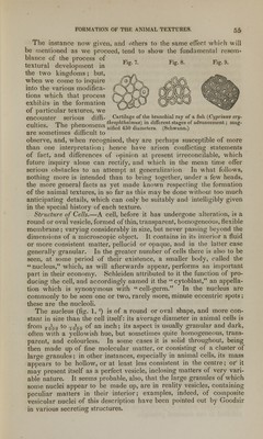
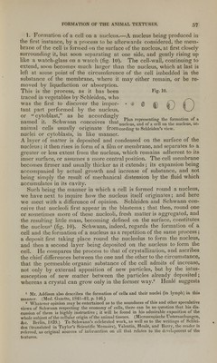
![substance of the stone or shell of most fruits (fig. 5 ;)] or they may assume the form of a slender fibre or band, single, double, or mul- tiple, running in a spiral manner along the in- sideofthe cavity, or forming a series of sepa- rate rings or hoops, as in spiral and annu- lar vessels (fig. 2,7). New matter may be absorbed or imbibed into the cells; or a portion of their altered and elaborated con- tents may escape as a secretion, either by transudation through the cell-wall, or by rupture or absorption of the membrane. Lastly, in certain circumstances, cells may be wholly or partially removed by absorp- tion of the membrane. Sections of cells st.-ongthened by 5. Cells may produce or generate new internal matter irregularly deposi- cells. The mode in which this takes place ted; the shaded portion indicates iU be immediateiy considered, in speak- the remaining cavities: a, cells from . . . J ' i the gritty centre ©f the pear ;&, cells ing ol the Origin ot animal Cells, from the stone of the plum.] FORMATION OF THE ANIMAL TEXTURES. Passing now to the development of the animal tissues, it may first be remarked generally, that in some instances the process exhibits an obvious analogy with that which takes place in vegetables; certain of the animal tissues, in their earlier conditions, appearing in form of a congeries of cells almost entirely resembling the vegetable cells, and, in their subsequent transformations, passing through a series of changes in many respects parallel to some of those which occur in the progress of vegetable development. Cartilage affords a good example of this. Figures 6 and 7, are magnified representations of cartilage in its early condition; and whoever compares them with the appearance of vegetable cells, shown in figures 1 and 2, must at once be struck with Fig.fi. the resemblance. Figs. 8 and 9, show the subsequent changes on the primary cells of cartilage; the parietes are seen to have become thickened by deposit of fresh material, the spaces within the cells, are consequently diminished, while the mass between the cavities is in- creased. Now this change seen to oc- cur in the cartilage cells, though there may be a question as to the precise mode (,„„,•„ - . ... t., - i'1 which it is brought about, may very Section of a branchial cartilage of a/••l 1 , ° . , . ' . . j ,olJ Tadpole, showing the early condition tairv be compared with the thickening of the cells; magnified 450 diameters.of the sides of the vegetable cells, which (Schwann.) takes p|ace when they gre converted into the woody and other hard tissues. Again, in most cartilages the cells increase in number as they diminish in size, new ones&bein«» formed within the old, as happens in many vegetable structures.](https://iiif.wellcomecollection.org/image/b21148867_0044.jp2/full/800%2C/0/default.jpg)




 No text description is available for this image
 No text description is available for this image
 No text description is available for this image