On the archetype and homologies of the vertebrate skeleton / by Richard Owen.
- Richard Owen
- Date:
- 1848
Licence: Public Domain Mark
Credit: On the archetype and homologies of the vertebrate skeleton / by Richard Owen. Source: Wellcome Collection.
Provider: This material has been provided by King’s College London. The original may be consulted at King’s College London.
123/238 (page 111)
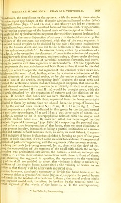
![]11 H 'J ■■(Si ■i jll 1. fti 'm . -'•D -t' I n- « ta u* ai t le correspondence with the scapular, or occipito-heeinal arch, is furtlier id out by the presence oiappendages which freely diverge Ironi it, but evelopiuent of these appendiiges has not been observed to extend beyond second phase, marked by vegetative multiplication of the simple ray, tly attached to the arch itself. The lepidosiren offers the simplest con- 1 of such ‘ diverging appendage ’ in the single slender bony piece con- ;d with the element 40*. Cuvier and other ichthyologists cite a series ages of this kind of development of the hyoidean appendage from a si- ^imple beginning up to a 30-fold repetition of the single ray {Flops); ;he ‘ brauchiostegal ’ rays have been found in much greater numbers in in fossil fishes. Like the ‘ pectoral ’ rays, they support a duplicature of brane, which plays freely backw’ards and forwards, reacting upon the ent medium, and forming, in short, a cephalic fin, but with its powers iftAstricted and adjusted, as to propel the water through the branchial cham- Jii .of the fish, instead of driving the fish through the water ; in which latter 'D on, indeed, the occipital appendages (pectoral fins)in most osseous fishes land do perform but a very small share. I we next proceed to compare the frontal segment, N iii and II in, dis- vbered as above described from the parietal vertebra, and, by the separa- of the sutures, from the bones terminating the skull anteriorly, we shall ra neural arch (fig. 3) closely repeating the characters of that of the oc- n.'i.al vertebra. The centrum is sometimes represented simply by the forward flsion of ossification of the basisphenoid (u), which I regard as the ho- jfpe of the ossification of the capsule of the notochord beneath the cen- wsof the anterior trunk-vertebrae in the silurus ; sometimes, also, of a di- ;t superincumbent symmetrical ossicle (o', fig. 5), answering to the rudi- Ltal (central part of the) body of the atlas supported by the inferior bony inthesilurus. This more complex condition of the centrum of the frontal «4ebra is well-seen in the sword-fish. The bones 10,10, which directly rest rjiio', when it exists, which defend the sides of the prosencephalon, and 13 ;.jh are either grooved by the optic nerves, or have those nerves perforating ifibro-cartilaginous membrane close to the margin of the bone (10) from :;;h it is continued, are obviously the neurapophyses. They are, however, 11; inasmuch as the segment of the brain to which they relate is of inferior in bony fishes : and they are still smaller in comparison with the spine • which is enormously expanded, in relation to its accessory functions as chief contributor to and protector of the orbits. The bones 12, wedged veen the neurapophyses and spine, affording an articular surface to the _ I i.dnial piece of the haemal arch, and developing a transverse process for «ular attachments, are the parapophyses. The bones (17) have as little ntial connection with the typical neural arch above demonstrated, as the ■es 18, la had with the corresponding arch of the parietal vertebra: and r more peculiar form in relation to the ball which they protect, and their able histological condition in the vertebrate series, have not only prevented r ever being mistaken for parts of cranial vertebrae, but have led to the •osite extreme of excluding them altogether from the bones of the skull, b which they are as much entitled to rank as the petrosal (10) or the 3inal(n); but always in the category of sense-capsules or ‘ splanchno- letal ’ pieces. n regard to the inferior arch of the frontal segment, the subdivision of its stituent elements, in subserviency to its special functions, is carried to as i at an extent as in that of the parietal segment. I regard the four over- I ping and closely-connected pieces from the upper joint {-mi) to the lower * Hunterian Lectures on Vcrtclimla. p. 79, fig. 27, 37.](https://iiif.wellcomecollection.org/image/b21307830_0123.jp2/full/800%2C/0/default.jpg)