Observations on the anatomy of Nycticebus tardigradus / by St. George Mivart and James Murie.
- St. George Jackson Mivart
- Date:
- [1865]
Licence: Public Domain Mark
Credit: Observations on the anatomy of Nycticebus tardigradus / by St. George Mivart and James Murie. Source: Wellcome Collection.
Provider: This material has been provided by The Royal College of Surgeons of England. The original may be consulted at The Royal College of Surgeons of England.
10/18 (page 248)
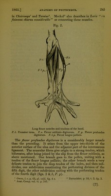
![other to the fourth digit in the right hand, but in the left hand to the fifth and index. This muscle, being quite single in our specimen, differs from that described by Meckel1 as existing in Loris. The extensor ossis metacarpi-pollicis shows no trace of subdivi- sion in its tendon, nor any indication that it really includes (as im- plied in the memoir) the extensor primi internodii. The extensor carpi ulnaris arises by two distinct heads, one from the posterior and lower surface of the external condyle, the other (fully half an inch broad) from the posterior surface of the ulna. These heads unite at an acute angle, and give origin to a tendon which has the usual insertion. Muscles of the Abdomen.—As regards the psoas and iliacus, we were uuable to determine satisfactorily their precise limits and sub- divisions ; nevertheless we are certain that neither the conditions described by S. Van der Kolk and W. Vrolik, nor those given by Meckel2 as existing in Loris, correspond with those in our specimen. The most internal portion, far from being “ la plus forte,” is the most slender. It is undoubtedly the psoas parvus, and arises by fleshy fasciculi from the sides of the bodies of the second and third lumbar vertebrae, hut very soon becomes entirely tendinous. Its long thin tendon, having an aponeurotic extension proceeding from the inner border, goes towards the pelvis, is closely applied to the muscle beneath, and finally inserted into the ilio-pectineal eminence imme- diately above the acetabulum. In Tarsius3 the tendon of the psoas parvus bifurcates. The large muscular mass beneath the above tendon appears to re- present the iliacus. It arises, however, from the sides of the bodies of the lumbar vertebrae below the third, and from the front of the sacrum (being separated from the pyriformis at its origin by the sacral plexus), but it has no origin from the ilium ; the insertion is normal. Another large muscle, which we suppose must be considered as the psoas macjnus, arises behind the last described (the ventral surface of the body being towards the observer), from the bodies and trans- verse processes, at their bases, of the two last dorsal and six upper lumbar vertebrae, and is inserted as usual. At its origin it has nu- merous tendons interspersed in its muscular substance, and is closely connected with the next to be described. This muscle, which we provisionally call *quadratus hmborum, arises from the two transverse processes of the lumbar vertebrae, from about the fourth downwards, and is inserted into the crest of the ilium immediately above what appeared to be the scansorius. Above it is so closely connected with the psoas magnus that the cor- rect determination of their limits requires further examination. The superficial abdominal muscles present nothing worthy of re- mark, excepting the rectus. This is continued forward as a narrow band on each side of the sternum, parallel with and about one-fifth 1 Anat. C6mp. vol vi. p. 324. 2 Loc. cit. p. 374. 3 Burrrieister, p. 67, t. 4. fig. 2. no. 1. [10]](https://iiif.wellcomecollection.org/image/b22352090_0012.jp2/full/800%2C/0/default.jpg)