Observations on the anatomy of Nycticebus tardigradus / by St. George Mivart and James Murie.
- St. George Jackson Mivart
- Date:
- [1865]
Licence: Public Domain Mark
Credit: Observations on the anatomy of Nycticebus tardigradus / by St. George Mivart and James Murie. Source: Wellcome Collection.
Provider: This material has been provided by The Royal College of Surgeons of England. The original may be consulted at The Royal College of Surgeons of England.
9/18 (page 247)
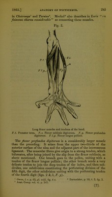
![In Cheiromys1 there is a very interesting intermediate condition. The arrangement much resembles that existing in Nycticebus; but the two muscles are not nearly so distinct. There is only one ten- don to the pollex, and the muscular fibres arising from the middle of the ulna join the part which answers to the flexor longus pollicis. The extensor communis digitorum and extensor minimi digiti ap- pear to be represented by only one muscle, which arises by a diminu- tive tendon from the external condyle of the humerus. The mus- cular fibres give origin to a tendon which soon becomes divided into two; the radial one of these again subdivides into four very fine tendons, one going to the index, another (the broadest) bifurcating, its branches going to the third and fourth digits respectively ; an- other (the third subdivision) goes to the fourth digit, and the last to the fifth digit. The ulnar main division of the common tendon goes to the fifth digit only. This last perhaps answers to the extensor minimi digiti, or that portion of the extensor communis recorded by Profes- sor Owen2 as existing in Cheiromys, and sending subsidiary tendons to the fourth and fifth digits. Fig. 4. Enlarged view of the palmar surface of the hand, to show the small muscles of the pollex and fifth digits; also the interossei and insertions of the lum- bricales. The extensor indicis is a very small muscle arising from the mid- dle of the ulna at its radial side, and from the interosseous mem- brane. It gives origin to two tendons, one going to the index, the 1 Owen, Trans. Zool. Soc. vol. v. p. 63, pi. xxiii. fig. 4. 2 Loc. cit. p. 62, pi. 23. fig. 2. no. 2 la. [9]](https://iiif.wellcomecollection.org/image/b22352090_0011.jp2/full/800%2C/0/default.jpg)