Licence: Public Domain Mark
Credit: Researches in embryology. (First series) / by Martin Barry. Source: Wellcome Collection.
Provider: This material has been provided by the Royal College of Physicians of Edinburgh. The original may be consulted at the Royal College of Physicians of Edinburgh.
20/64 (page 316)
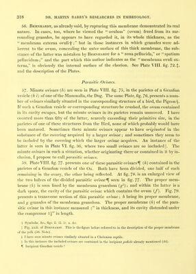
![just before the ovum leaves the ovary, this membrane, previously so delicately thin, becomes perfectly distinct and very thick; and that the chorion, imbibing fluid into its interior, becomes somewhat distended, so that a minute space is visible between the membrana vitelli and the chorion. This thickening of the proper membrane of the yelk, and the distention of the chorion, subsequently proceed much farther, as is proved by the state of ova found in the Fallopian tube. I find also, that the mem- brana vitelli is still visible, and has considerable thickness in minute ova met with in the uterus. This subject will be entered into more fully in a future paper. The true Chorion, a Structure superadded within the Ovary in the Class Mammalia. 52. The figures above referred to, in comparing the ovisac of Mammalia with that of Birds, Amphibia, and Fishes, present also up to a certain period a perfect analogy between the rudiments of the ovum itself in these four classes. We have seen in all, the germinal vesicle and its contents, as well as the yelk, and proper membrane of the yelk. Here, however, the analogy is terminated by the formation, within the ovary, in Mammalia, of a membrane to which there is no corresponding structure within the ovary in other Vertebrata. This membrane appears to be the true chorion. In the ovary of Birds, Amphibia, and Fishes, it is, I believe, allowed that there is no membrane formed external to the membrana vitelli-i-. The body therefore expelled from the ovary in these animals is not an ovum, but a yelk-hall. The subject will be illustrated by the following Table, showing the parts present (in a mature state) in the ovary of Mammalia on the one hand, and of Birds, &c. on the other:— Mammalia. Birds, some Amphibia^, and some Fishes:[. Germinal vesicle (c) and its contents. Yelk. d. Membrana vitelli, e. Chorion, f. Germinal vesicle (c) and its contents. Yelk, d. Membrana vitelli, e. ^Tunica granulosa, g^. §Retinacula, g-. Fluid, granules (g), and oil-like globules (*). § Membrana granulosa, g^. Ovisac, h. \ n . , , . Vascular tunic, i. / = Graafian vesicle, h t. Stroma, k. Peritoneal covering. I. Ovisac, h, . 1 1 7 • /'..>= Capsule, n, i vascular tunic, I. ] ^ Stroma, k. Peritoneal covering, /. > = Calyx, h i k I. t Professor Rathke, however, finds that in certain Fishes, not provided with an oviduct, the schaalenhaut is a production of the ovary. (Burdach's Physiologic, 1837, Band II. § 339.) t In other Amphibia and most of the osseous Fishes, the peritoneum does not enter into the formation of the calyx (47. Note). § To be described in Part II. of this Memoir (64. 80. 72.).](https://iiif.wellcomecollection.org/image/b21972138_0020.jp2/full/800%2C/0/default.jpg)