Text-book of human physiology : including histology and microscopical anatomy, with especial reference to the practice of medicine / by L. Landois.
- Date:
- 1904
Licence: In copyright
Credit: Text-book of human physiology : including histology and microscopical anatomy, with especial reference to the practice of medicine / by L. Landois. Source: Wellcome Collection.
Provider: This material has been provided by the Royal College of Physicians of Edinburgh. The original may be consulted at the Royal College of Physicians of Edinburgh.
1017/1084 (page 1001)
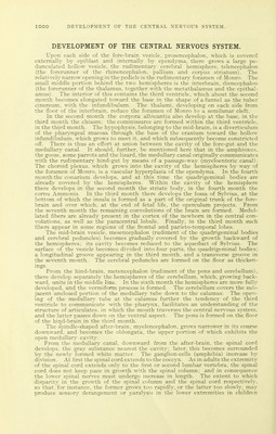
![should be kept in mind. The tactile nerves of the fetus are capable of executing reflex movement, for example on pressure upon the palpable fetal parts. The first indications of the muscles appear upon the back in the second month; in the fourth month, they become reddish. The first appreciable fetal movements occur about the middle of pregnancy; these are reflex, as they are observed also in acephalous fetuses. It is noteworthy that, in the early periods of develop- ment, the central nervous system has no functional influence upon the vital processes, having no sensory, or motor, or trophic (morphogenetic) function, as has been demonstrated by the extirpation-experiments of Alf. Schapee. The spinal ganglia develop from a special band, situated on each side of the medullary canal, and forming the direct connection between this and the epi- dermis. The spina] ganglia are the nuclei of origin of the sensory nerves, whence a communication with the spinal cord is established and the peripheral nerve-trunks grow in a centrifugal direction. The nerves of special sense also /. s. iv: V Fig. 394.—Development of the Eye: /, Invagination of the lenticular sac (i) into the primary optic vesicle (P); e, epiderm; m, mesoblast; 77, the invaginated primary optic vesicle newed from below; optic nerve; a the outer, i the inner layer of the invaginated vesicle; i, lens; 777, the same formation in longitudinal section; 7F, further development; f, corneal epithelium; c, cornea; m, capsulopupillary membrane; i, lens; a, central artery of the retina; i, sclera; cA, choroid; p, pigment-epithelium of the retina; r, retina; F, per- sistent vestige of the pupillary membrane. (Diagrammatic.) grow from the periphery into the central organ. The motor nerve-roots grow from the rudimentary ganglia in the central organ (neuroblasts) into the periphery. At first the nerves are non-medullated. Human embryos four weeks old possess spinal ganglia, anterior roots and in part the tmnks of the spinal nerves, whereas the posterior roots are absent. The ganglia of the fifth, seventh, eighth, ninth, and tenth cranial nerves, and in part their origins, are present; on the other hand. His failed to find the first, second, third, and twelfth cranial nerves, as well as the sympathetics. Fetuses with absence of the spinal cord show that the posterior roots present and the sensory nerves originate from the spinal ganglia. In the new-bom the cranial motor nerves and the auditory are already provided with medullary sheaths; the others are not. Their envelopment pro- gresses peripherally. In the peripheral spinal nerves the formation of the medul- lary sheath does not take place before the second and third years. The sympathetic ganglia of the viscera make their way from the sympa- thetic cord into the organs. DEVELOPMENT OF THE ORGANS OF SPECIAL SENSE. Eye.—The primary optic vesicle grows out to the external covering of the head (epiblast) and then becomes invaginated into itself from before backward (as has been seen to take place in human embryos four weeks old), so that the pedunculated vesicle has acquired the shape of an egg-cup (Fig. 394, /). The interior of this cup, the subsequent cavity of the eye, is now called the secondary optic vesicle. The portion of the original vesicle that has undergone invagina- tion, namely the anterior convex portion, now made concave, becomes the retina {IV, r); the posterior portion of the vesicle becomes the pigmented choroidal (retinal) epithelium (IV, p). The pedicle is the subsequent optic nerve. The invagination of the primary optic vesicle takes place, however, not exactly accord- ing to this simple plan; but there is formed below on the egg-cup-shaped structure](https://iiif.wellcomecollection.org/image/b21981504_1017.jp2/full/800%2C/0/default.jpg)