Text-book of human physiology : including histology and microscopical anatomy, with especial reference to the practice of medicine / by L. Landois.
- Date:
- 1904
Licence: In copyright
Credit: Text-book of human physiology : including histology and microscopical anatomy, with especial reference to the practice of medicine / by L. Landois. Source: Wellcome Collection.
Provider: This material has been provided by the Royal College of Physicians of Edinburgh. The original may be consulted at the Royal College of Physicians of Edinburgh.
964/1084 (page 948)
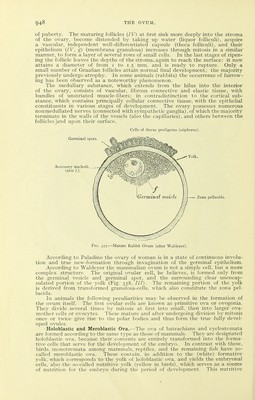
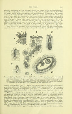
![of puberty. The maturing follicles {IV) at first sink more deeply into the stroma of the ovary, become distended by taking up water (liquor folliculi), acquire a vascular, independent well-differentiated capsule (theca folliculi), and their epithelium {IV, g) (membrana granulosa) increases through mitosis in a similar' manner, to form a layer of several rows of small cells. In the last stages of ripen- ing the follicle leaves the depths of the stroma, again to reach the surface: it now attains a diameter of from i to 1.5 mm. and is ready to rupture. Only a small number of Graafian follicles attain normal final development; the majority previously undergo atrophy. In some animals (rabbits) the occurrence of furrow- ing has been observed as a noteworthy phenomenon. The medullary substance, which extends from the hilus into the interior of the ovary, consists of vascular, fibrous connective and elastic tissue, with bundles of unstriated muscle-fibers; in contradistinction to the cortical sub- stance, which contains principally cellular connective tissue, with the epithelial constituents in various stages of development. The ovary possesses numerous nonmedullated nerves (connected with sympathetic ganglia), of which the majority terminate in the walls of the vessels (also the capillaries), and others between the follicles ]and upon their surface. Cells of discus proligerus (uophorus). Ac, pellucida. Fig. 357.—Mature Rabbit Oram (after Waldeyer). According to Paladino the ovary of woman is in a state of continuous involu- tion and true new-formation through invagination of the germinal epithelium. According to Waldeyer the mammalian ovum is not a simple cell, but a more complex structure. The original ovular cell, he believes, is formed only from the germinal vesicle and germinal spot, and the surrounding clear unencap- sulated portion of the yolk (Fig. 358, ///). The remaining portion of the yolk is derived from transformed granulosa-cells, which also constitute the zona pel- lucida. In animals the following peculiarities may be observed in the formation of the ovum itself. The first ovular cells are known as primitive ova or ovogonia. They divide several times by mitosis at first into small, then into larger ova- mother cells or ovocytes. These mature and after undergoing division by mitosis once or twice give rise to the polar bodies and thus form the true fully devel- oped ovvdes. Holoblastic and Meroblastic Ova.—The ova of batrachians and cyclostomata are formed according to the same type as those of mammals. They are designated holoblastic ova, because their contents are entirely transformed into the forma- tive cells that serve for the development of the embryo. In contrast with these, birds, monotremata among mammals, reptiles, and the remaining fish have so- called meroblastic ova. These contain, in addition to the (white) formative yolk, which corresponds to the yolk of holoblastic ova, and yields the embryonal cells, also the so-called nutritive yolk (yellow in birds), which serves as a source of nutrition for the embryo during the period of development. This nutritive](https://iiif.wellcomecollection.org/image/b21981504_0964.jp2/full/800%2C/0/default.jpg)