Text-book of human physiology : including histology and microscopical anatomy, with especial reference to the practice of medicine / by L. Landois.
- Date:
- 1904
Licence: In copyright
Credit: Text-book of human physiology : including histology and microscopical anatomy, with especial reference to the practice of medicine / by L. Landois. Source: Wellcome Collection.
Provider: This material has been provided by the Royal College of Physicians of Edinburgh. The original may be consulted at the Royal College of Physicians of Edinburgh.
987/1084 (page 971)
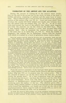
![THE EMBRYONIC HEART. also this projects free, with the formation of the tail-fold (S) and the hind-gut (d), to which the posterior intestinal portal leads. The em- bryonal body thus communicates with the germinal vesicle by means of a pedicle that is as first wide open. This pedicle is known as the omphalomesenteric or vitellointestinal duct. The saccular vesicle attached to it is designated in mammals the umbilical vesicle (VII, N), while the analogous much larger sac in birds, which contains nourishment from the yellow yolk, is known as the yolk-sac. Toward the end of the third month of pregnancy the entodermal lining of the human umbilical vesicle develops genuine liverlike glandular tissue. The omphalomes- enteric duct becomes in its further course narrower and finally is oblit- erated in the chick on the fifth day. Where the duct is inserted into the abdominal wall there results the abdominal umbilicus ; where it is inserted into the primitive gut there results the intestinal navel. On the ventral surface of the fore-gut and the hind-gut there are points where the mesoderm is wanting, and where, therefore, the epiblast and the entoblast come in contact. These are known as the pharyngeal and the cloacal membrane. The openings for the formation of the oral and the anal orifices are later found in these situations. Even before this process of constriction takes place the primitive heart develops from that portion of the splanchnopleure that is in con- tact below with the fore-gut, in the chick at the conclusion of the first day as a rhythmically moving point {^(niypr] -/fjouni'^rj of Aristotle, punctum saliens); in mammals however, much later. The heart (Fig. 376, VI) develops as a cellular, hollow, bladderlike bud of the splanchnopleure (originally as a paired structure). Its cavity soon dilates and it grows into the coelom suspended from a mesenterj^-like duplicature (mesocar- dium); that part of the coelom situated in the vicinity of the heart is now designated the cardiac fossa (fovea cardiaca). The heart acquires a longitudinal tubular form, with its aortic portion directed anteriorly and its venous portion directed posteriorly. It then undergoes a mod- erate 5-shaped curvature (Fig. 384, i). From the middle of the second day the heart in the chick beats regularly, about 40 times per minute. At the anterior (aortic) extremity of the heart,' the aorta originates from the bulbus aortae; it bends forward, and, dividing into two arches (primitive aortas), it curves beneath the brain-vesicles and descends posteriorly in front of the primitive vertebrae. Both primitive aortas originally terminate blind at the caudal extremity of the embryo. Opposite the omphalomesenteric duct each primitive aorta in chicks gives off one, in mammals several (in the dog 4 or 5) omphalomesenteric arteries (Fig. 376, VI, Ao) which divide within the mesoblast upon the yolk-sac, or the umbilical vesicle, into a rich network of vessels. These unite and, passing backward (in birds arising from the terminal sinus of the subsequent terminal vein of the area vasculosa), form omphalomes- enteric veins (Vo), which ascend on the duct and empty into the two venous trunks of the heart by means of two branches. Thus the first or primitive circulation is completed. Its pur- pose is to convey nutritive material for growth and oxygen to the embryo. The latter, in birds, passes through the porous shell of the egg from the air; the first is supplied by the yolk-sac until the end of the incubation. In mammals both are supplied to the ovum from the vessels of the uterine mucosa. In birds, on account of the consumption](https://iiif.wellcomecollection.org/image/b21981504_0987.jp2/full/800%2C/0/default.jpg)