Lectures on the comparative anatomy of the placenta : first series / delivered before the Royal College of Surgeons of England, June, 1875, by Wm. Turner.
- William Turner
- Date:
- 1876
Licence: Public Domain Mark
Credit: Lectures on the comparative anatomy of the placenta : first series / delivered before the Royal College of Surgeons of England, June, 1875, by Wm. Turner. Source: Wellcome Collection.
Provider: This material has been provided by The Royal College of Surgeons of England. The original may be consulted at The Royal College of Surgeons of England.
35/132 (page 35)
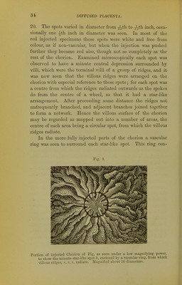
![sisted of a double row of capillaries, which passed from the capillaries of one villous ridge to those in the next adjacent ridges, so that, the vessels in the whole series of ridges radiating from any given spot being connected together, a vascular ring was formed. But it was particularly observed that this ring was not formed of vessels passing between the central ends of the ridges, but a little to their outer side, so that the ends were enclosed by the vascular ring. The existence of small circular spots whiter than the sur- rounding parts of the chorion was recognised so far back as 1781 by John Hunter\ Von Baer gave a careful description^ of their star-like arrangement, and described and figured their vascularity; more particularly did he describe the relations of their capillaries to the branches of the umbilical vein, so that if a blue injection be introduced into the umbilical vein, and a red into the artery, these spots would appear as blue stars on a red ground. Eschricht also examined these spots ^ and arrived at a similar conclusion as to their relation to the umbilical vein. M. Flourens described* these spots as small discs, which are, he states, a variety of the multiple form of placenta. He would obviously associate them therefore with the foetal cotyledons of a ruminant, v But this mode of regarding them is quite erro- neous, for the centre of each spot is non-villous, and when the chorion is in situ, as I shall immediately point out, is in contact with a feebly vascular part of the uterine mucosa and not with the highly vascular crypts. Each end of the chorion, for nearly three inches from the pole, had a smooth non-villous surface, and though possessing considerable vascularity was not so vascular as the villous part of the chorion. Hence the chorion of the pig is not uniformly villous, but the villi, as was indeed known to von Baer, arc distributed over the central and not the polar regions of the 1 Essays and Observations, edited by Prof. Owen, Vol. i. p. 198. Loudon, 18G1. 3 Untersuchungen iiber die Gefcissverhindung zwischen Mutter und Frucht, p. 9, Fig. ], 2. Leipzig, 1828. De orrjanis qtuc respirationi et nutritioni fcetus mammaliuni inscrviunt, p. 36. Ilajniee, 1837. ■» Conrs sur la Generation, rccueilli ct pulli6 par M. Descliamps, p. 147. Paris, 183G.](https://iiif.wellcomecollection.org/image/b22294478_0037.jp2/full/800%2C/0/default.jpg)