Lectures on the comparative anatomy of the placenta : first series / delivered before the Royal College of Surgeons of England, June, 1875, by Wm. Turner.
- William Turner
- Date:
- 1876
Licence: Public Domain Mark
Credit: Lectures on the comparative anatomy of the placenta : first series / delivered before the Royal College of Surgeons of England, June, 1875, by Wm. Turner. Source: Wellcome Collection.
Provider: This material has been provided by The Royal College of Surgeons of England. The original may be consulted at The Royal College of Surgeons of England.
54/132 (page 54)
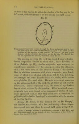
![54i surface of the chorion to within two inches of its free end in the left cornu, and nine inches of its free end in the right cornu. Fig. 10. Diagrammatic transverse section through the foetus and membranes to show the relations of the allantois to the abdominal aspect of the foetus, and the relations of the amnion to the chorion. E. embryo, ch. the vUlous chorion, al. the allantois, the letters are in the sac. The dotted line am. am. represents the amnion, the letters are in the sac. The amnion investing the cord was studded with yellowish- brown corpuscles, similar to those that I have described in Orca gladiator (p. 23); similar corpuscles were scattered in considerable numbers over the amnion covering the allantois, and a few were seen on the amniotic lining of the chorion. But in addition, numerous dull Avhite corpuscles were found, some of which were slender rods, from -J^th to ^^th inch long, and arranged end to end like the links of a chain; whilst others were globular, like small shot. The rods were most numerous on the abdominal half of the cord, the globules at and near its bifurcation. These corpuscles were, like those of a yellowish- brown colour, covered by the amnion. When examined micro- scopically they were found to be composed of crowds of squa- mous epithelial cells, so that they resembled in structure the yellowish-white bodies developed in connection with the amnion of the Cow (p. 25). Manis.—In Mams, as was pointed out by Dr Sharpey\ the chorion was covered with fine reticulating villous ridges, interrupted here and there by round bald spots, giving it an 1 Quoted in Huxley's EZcjncnt.'? o/ Comparative Anatomy,]). 112, 1864; and with additional details in my Memoir on the Placentation of the Sloths in Trans. Hoy. Snc. Edinh. 1873.](https://iiif.wellcomecollection.org/image/b22294478_0056.jp2/full/800%2C/0/default.jpg)