Rudiments of animal physiology : for use in schools, and for private instruction / by G. Hamilton.
- Hamilton, George, 1808-1885
- Date:
- 1839
Licence: Public Domain Mark
Credit: Rudiments of animal physiology : for use in schools, and for private instruction / by G. Hamilton. Source: Wellcome Collection.
Provider: This material has been provided by the Francis A. Countway Library of Medicine, through the Medical Heritage Library. The original may be consulted at the Francis A. Countway Library of Medicine, Harvard Medical School.
87/116 page 81
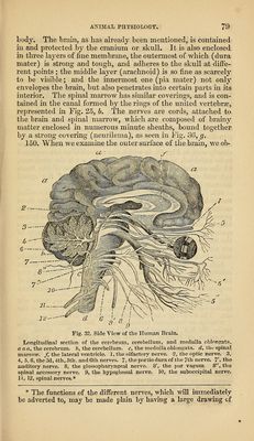
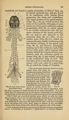
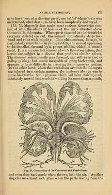
![examined, are found to consist of bundles of fibres,* that can be traced upwards into, and are found to be continuous with, similar fibres composing the brain and cerebellum. The upper portion of the spmal marrow ^ (Figs. 32, c, 83, /), which receives the ^ name of the medulla oblongata, is com- ' ^ posed, 1st, of two parts called the corpora ^jQ pyramidalia (Fig. 35, e), which appear to be chiefly continuous with the an- terior columns of the spinal cord (Fig. 36, a), and to i-un upwards to the cerebrum (Fig.'85, a); 2dly, of two :^ similar parts (Fig. 35,//), called the corpora olivaria, chiefly continuous with the lateral columns of the spinal cord (Fig. 36, 6), and likewise principally running up to the cerebrum; and, Sdly, of two other parts, called corpora resti- formia, behind the corpora olivaria, con- tinuous with the posterior spinal co- lumns (Fig. 36, c), and chiefly running to the cerebellum (Fig. 35, 6).t The two lobes of the cerebellum are also con- nected with each other by a part (Fig. 35, g) called the bridge of Varolius. * The interesting discovery has been made within these few years, by German anatomists, that these fibres, and all the fibres com- posing the nervous matter.are tubes filled with a fluid. The annexed sketch (taken from the British and Foreign Me- dical Review, No 12, in which a detailed account of this dis- covery is given), shows tlie Fig- 34. fibres of one of the nerves mag-nified, with the fluid contained in them escaping from their extremities. It is pro- bable that this discovery may be the means of throwing some light upon the functions of this hitherto little understood part of the animal frame, i* Sir C. Bell has described another part, which he calls the respi- ratory column, but it has not been generally admitted by pliysiologists. On separating the two corpora pyramidalia, the fibres can be seen very distinctly to cross from the right and left sides, and this is thought to be the reason Avhy palsy, from injury or disease of the one hemi- sphere of the brain, frequently afi'ects the opposite side of the body. % View of the base of the brain, anterior part of the spinal marrow, and attached nerves, a, Cerebrum; 6, cerebellum; c, spinal marrow; /, medulla oblongata. Fig. 33. Base of the Brain and Spinal Cord.:]:](https://iiif.wellcomecollection.org/image/b21056766_0087.jp2/full/800%2C/0/default.jpg)
 No text description is available for this image
 No text description is available for this image
 No text description is available for this image