The house fly Musca domestica, Linnæus : a study of its structure, development, bionomics and economy / by C. Gordon Hewitt.
- Hewitt, C. Gordon (Charles Gordon), 1885-1920.
- Date:
- 1910
Licence: In copyright
Credit: The house fly Musca domestica, Linnæus : a study of its structure, development, bionomics and economy / by C. Gordon Hewitt. Source: Wellcome Collection.
Provider: This material has been provided by London School of Hygiene & Tropical Medicine Library & Archives Service. The original may be consulted at London School of Hygiene & Tropical Medicine Library & Archives Service.
122/258 page 524
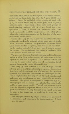
![524 0. GORDON HEWITT. channels to the mouth. Distally many of the channels unite; the resulting channels all converge and run into the mouth. The anterior border of the oral aperture is occupied by the mandibular sclerite (m. s.), and the posterior border is bounded by a lingual-like process (I.) that is bilobed at its anterior extremity. Cephalo-pharyngeal sclerites (PI. 30, fig. 4).—The sclerites associated with the cephalo-pharyngeal region are rather similar to those of the second larval instar; they are, however, of a more solid and of a thicker character. Between the oral lobes is seen the median uncinate mandibular sclerite (m. s.). The homology of this sclerite is obscure. Lowne regarded it as being the labrum ; some authors con- sider that it represents the fused mandibles. As we know at present so little of the comparative embryology of these larvae it will be best to retain the name by which it is generally known. The basal extremity of the mandibular sclerite is broad, and at each side a dentate sclerite (d. s.) is articulated by means of a notch in the side of the mandibular sclerite, the function of which has been shown already in describing the muscles. The mandibular sclerite articulates posteriorly with the hypostomal sclerite (h. s.). This consists of two irregularly-shaped lateral portions united by a ventral bar of chitin; it is anterior to this bar of chitin that the salivary duct opens into the front of the pharynx. The sides of the hypostomal sclerite articulate with two processes on the anterior edge of the lateral pharyngeal sclerites (I. p.). The lateral pharyngeal sclerites or plates i*ecall the shape of the fulcrum of the adult fly. Each is wider posteriorly than anteriorly, and the posterior end is deeply incised; at the base of this incision the nerves and tracheae which supply the interior of the pharynx enter. The lateral sclerites vary in thickness, as will be seen in the figures of the sections of the pharynx. They are united dorsally at the anterior end by a dorsal sclerite (d. p. s.), and ventrally they are continuous with the floor of the pharynx. The pharynx (PI. 3], figs. 17 and 18) in certain respects is](https://iiif.wellcomecollection.org/image/b21357110_0122.jp2/full/800%2C/0/default.jpg)
 No text description is available for this image
 No text description is available for this image
 No text description is available for this image