Licence: Public Domain Mark
Credit: Heart / by John Reid. Source: Wellcome Collection.
Provider: This material has been provided by the Royal College of Physicians of Edinburgh. The original may be consulted at the Royal College of Physicians of Edinburgh.
14/46 page 14
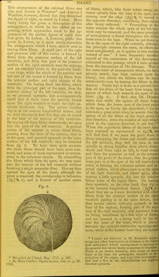
![in the heart of the ox and horse after a little dissection. The following description is drawn up from numerous dissections of tliese parts made on the human heart. The tendinous ring surrounding tlie aortic opening is stronger and tliicker than that surrounding the orifice of the puhnonary artery. Both of them are stronger than the auriculo-ventricular rings. Each of the arterial rings appears as if composed of three semilunar portions placed on tlie same ])lane, the convexities of which are turned towards the ventricles and the concavities to- wards the vessels (fig. 1, a a).* Each of Fig. 2. Appearance of tendinous ring at the origin of tlie puhnon/iry artery. In slitting open the artery, one of the three projecting extremities of tlie ten- dinous ring has been divided. these semilunar portions has its projecting extre- mities intimately blended at their terminations with the corresponding projecting extremities of those next to it, (Jig. 1, h b,) so that the three form a complete circle, with three trian- gular portions projecting from its upper edge. The semilunar portions approach fibro-carti- lage in their structure, and have the intervals left between their convex edges filled with a texture more decidedly fibrous, (fig. 1, and which is considerably weaker than the se- milunar portions, more particularly on the left side of the heart.f The thinness of the ten- dinous structure filling up these intervals has led some anatomists erroneously to describe these portions of the heart as protected only by the two serous membranes. The right ten- dinous zone is broader than the left and very thin, particularly at its inner margin, at which part in both sides of the heart it assumes mote of the tendinous than of the fibro-cartilaginous structure. These tendinous rings are placed obliquely from without inwards and from above downwards, so that the outer edge is on a plane superior to the inner. The sigmoid valves are attached to the inner edge of the upper surface, (fig. 2, Oy) and the tendinous fibres placed in the fixed margins of these valves contribute to the thickening of the ring at this part; the middle coat of the arteries is connected to the outer edge of the same surface, and to the an- terior part of the projecting extremities, (fig. 2, b;) while the muscular fibres of the ven- tricles (fig. 1,/; fig- 2, are attached to the lower surface of the projecting portion of the convexity, and to tlie lower margin of the fibrous tissue filling up the space between the convexities of the projecting ends, (fig. • These tendinous festoons are represented stronger in the woodcut than tlicy are naturally. t These intervals are occupied by muscular ribres in the heart of the ox and horse. Puhnonary artery slit open at its origin, it- miinuil membrane stripped off, and two of the sigmoid valves completely removed. a a a, tendinous festoons. b b, muscular fibres of the right ventricle. c c c, middle fibrous coat of the artery after the internal serous membrane has been stripped off. g, small portion of one of the semilunar valves left to shovv its attachment to the inner edge of the upper surface of the tendinous festoon. 2, d.) There is, however, this difference between the right and left arterial openings with respect to the attachment of the muscular fibres;—on the right side the muscular fibres arise from the projecting portion of the con- vexity of the whole three tendinous festoons, (fig. 3, c, c,) while in the left side the mus- cular fibres are attached only to one and part of a second, (fig. 4, b b,) as the larger lip of the mitral valve (fig-4, a) is suspended](https://iiif.wellcomecollection.org/image/b21908503_0016.jp2/full/800%2C/0/default.jpg)