Lectures on surgical pathology : delivered at the Royal College of Surgeons of England / by James Paget ; rev. and ed. by William Turner.
- James Paget
- Date:
- 1865
Licence: Public Domain Mark
Credit: Lectures on surgical pathology : delivered at the Royal College of Surgeons of England / by James Paget ; rev. and ed. by William Turner. Source: Wellcome Collection.
Provider: This material has been provided by the Augustus C. Long Health Sciences Library at Columbia University and Columbia University Libraries/Information Services, through the Medical Heritage Library. The original may be consulted at the the Augustus C. Long Health Sciences Library at Columbia University and Columbia University.
708/742 page 710
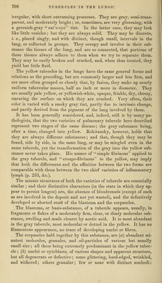
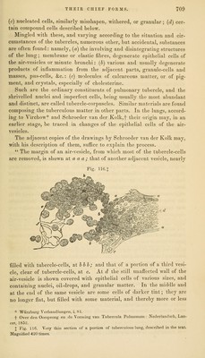
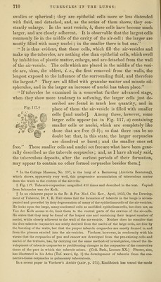
![swollen or spherical; they are epithelial cells more or less distended with fluid, and detached, and, as the series of them shows, they con- stantly enlarge. In the next vesicle, h, these cells have become much larger, and are closely adherent. It is observable that the largest cells commonly lie in the middle of the cavity of the air-cell: the larger are mostly filled with many nuclei; in the smaller there is but one. It is thus evident, that these cells, which fill the air-vesicles and make up the tubercles, are nothing else than epithelial cells, which swell by imbibition of plastic matter, enlarge, and are detached from the wall of the air-vesicle. The cells which are placed in the middle of the vesi- cle are, thus, the oldest, i. e., the first removed from the walls, the longest exposed to the influence of the surrounding fluid, and therefore the largest.* They are all filled with granular matter and minute oil- spherules, and in the larger an increase of nuclei has taken place. If tubercles be examined in a somewhat further advanced stage, when they show more tendency to softening, the larger cells just de- scribed are .found in much less quantity, and in place of them the air-vesicle is filled with smaller cells [and nuclei]. Among these, however, some larger cells appear (as in Fig. 117, a) containing smaller cells or nuclei, which are completely like those that are free {h b) ; so that there can be no doubt but that, in this state, the larger corpuscles are dissolved or burst; and the smaller ones set free. These smaller cells and nuclei set free are what have been gene- rally described as the tubercle corpuscles; and, as I have already said, the tuberculous deposits, after the earliest periods of their formation, may appear to contain no other formed corpuscles besides them. J * In the College Museum, No. 297, is the lung of a Benturong (Arctictis Benturong), which shows, apparently very well, this progressive accumulation of tuberculous matter from the walls to the centres of the air-cells. f Fig. 117. Tubercle-corpuscles : magnified 420 times and described in the text. Copied from Schroeder van der Kolk. X In an elaborate paper in the Br. & For. Med. Chi. Rev., April, 1855, On the Develop- ment of Tubercle, Dr. C. R. Hall states that the formation of tubercle in the lungs is accom- panied and preceded by fatty degeneration of many of the epithelium-cells of the air-vesicles. He looks upon the large, many-nucleated cells as modified epithelium-cells, but does not, as Van der Kolk seems to do, limit them to the central parts of the cavities of the air-cells. He states that they may be found of the largest size and containing their largest number of nuclei, while closely adherent to the wall of the air-vesicle. Neither does he consider that the free tubercle corpuscles are solely derived from the nuclei of the large cells, set free by the bursting of the walls, but that the proper tubercle corpuscles are mostly formed in and from the plasma exuded into the air-vesicles. Virchow, however, in conformity with his views that the corpuscles of pus and cancer are developed from the pre-existing cells and nuclei of the textures, has, by carrying out the same methods of investigation, traced the de- velopment of tubercle corpuscles to proliferating changes in the corpuscles of the connective tissue of the part in which the tubercle arises. (Cell. Path., Lecture XX.) Ffirster, also, has illustrated in his Atlas (Taf. xxxvi, fig. 1) the development of tubercle from the con- nective-tissue corpuscles in pulmonary tuberculosis. In a recent paper in Virchow's Archiv (xxiv, p. 571), Rindfleisch has traced the mode](https://iiif.wellcomecollection.org/image/b21211267_0708.jp2/full/800%2C/0/default.jpg)