Report on the outbreak of plague at Sydney [1900-1907] / by J. Ashburton Thompson, Chief Medical Officer of the Government and President of the Board of Health.
- New South Wales. Department of Public Health
- Date:
- 1900-1908
Licence: In copyright
Credit: Report on the outbreak of plague at Sydney [1900-1907] / by J. Ashburton Thompson, Chief Medical Officer of the Government and President of the Board of Health. Source: Wellcome Collection.
Provider: This material has been provided by London School of Hygiene & Tropical Medicine Library & Archives Service. The original may be consulted at London School of Hygiene & Tropical Medicine Library & Archives Service.
75/452 page 69
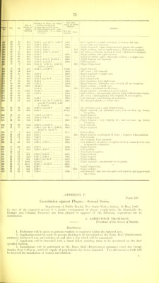
![6^ Case 240. May 20. M., ait 45. Duration of illness, 12 hours. Body very obese. A few petechiaj were found on the trunk and limbs. Thorax.—Lungs somewhat engorged. Heart.—The walls of the heart, especially the left ventricle, were of soft consistence, and mottled from early fatty degeneration. The cavities of the organ contained a small quantity of dark fluid blood. Valves were healthy. Abdomen.—Liver markedly enlarged, and the seat of extensive fatty degeneration. Spleen enlarged, of normal consistence. On section of a dark red colour. Trabecule obscured. Kidneys somewhat enlarged and of soft consistence. On section the capsule stripped readily. The cortex was paler than usual and mottled throughout with numerous pin-point hremorrhages. iStomach showed evidence of chronic gastric catarrh, and also numerous recent submucous htemorrhages. Lymphatic Glands.— In the right femoral region one of the glands was found to be swollen slightly, and to be surrounded by a small hiemorrhagic zone. On section it had a mottled appearance. Microscopic examination of the spleen and lymphatic gland pulps showed numerous plague bacilli. S.J. Case 242. May 21. M., eel 17. Dwraiion of illness, 12 hoiws. External appearances.—A few petechia? on sides of chest and on back. Rigor mortis present. Lividity marked. The glands in both inguino-femoral regions were felt to be enlarged and shotty. Thorax.—Lungs somewhat engorged. No sub-jileural hemorrhages. Heart apparently healthy. A parti coloured clot filled the right auricle and ventricle. Abdomen.—Liver enlarged and slightly mottled. A few petechial htemorrliages beneath the capsule of Glisson. In one small area in the substance of and on the surface of the organ was a group of small pin- point yellowish-white bodies closely resembling miliary tubercles. Spleen enlarged and almost semi-diffluent. On section it was of a dark reddish-brown colour. Kidneys beyond being somewhat congested showed no abnormality. Stomach.—The mucous membrane was swollen, red, mamraillated, and at the cardiac end were numerous small submucous hiieraorrhages. Lymphatic Glands.—The glands in both inguino-femoral regions v\-ere found to be somewhat enlarged. In consistence they were harder than normal (resembling the condition seen in syphilitics). (_)ne of the femoral glands on the right side was somewhat redder than the rest, and on section were somewhat mottled. Microscopic Examination.—Films were made, both of the spleen pulp and of the jndp of the gland described above. No organisms resembling the plague bacillus were found in the spleen, but the film taken from the gland contained innumerable plague bacilli. S.J. Case 255. May 31. M., ml 55. Duration of illness, 15 lionrs. The body was that of a muscular and well-nourished man ; the face and neck deeply cyanosed ; the mouth contained some semi-digested food. No enlarged glands. Old pleuritic adhesions in both sides of chest, the large bronchi inflamed, and the lungs somewhat congested. Heart large. Left ventricle hyper- trophied ; heart much diseased. Coronary vessels slightly atheromatous. Dark clots in both sides. Liver in an advanced stage of cirrhosis. Kidneys granular. Stomacii contained a quantity of food ; its mucous coat in a condition of chronic inflammation. S|)]een enlarged ; softer than normal. This appeared to be a recent change, and was a little suggestive of plague. In the brain the arachnoid was thickened and opaque, witli some atrophy of the convolutions. A piece of the spleen was removed and handed to Dr. Tidswell, and, after examination, the case was reported as one of plague. G.H.T. Case 295. June 29. J/., cH. 55. Duration of illness, 70 hours. Body very emaciated. A swelling about the size of a hen's egg in the right groin. Numerous petechia^ on the trunk and limbs. On the right leg were two small vesicular papuke, which looked as though they had had their heads .scratched off. Thorax.—Both pleural sacs were entirely obliterated by old, tough, fibi'ous adhesions, the result of previous repeated attacks of pleurisy. Lungs.—At the apices of both lungs were old deposits of chronic fibroid tubercle, and in the upper lobe of the right lung was a somewhat more recent spread of miliary tubercle. The bronchi showed signs of chronic catari'hal inflammation. Heart.—There was considerable hypertrophy of the right ventricle. Both ventricles were distended by blood clot ; that on the right side was for the most part of a yellowish-white colour, and slightly adherent to the columns carnea-. On the left side the clot was dark-coloured and very crumbly. At the root of the aorta was some evidence of advanced atheroma ; otherwise the heart showed no abnormality. Abdomen.—There were no ecchymoses on the peritoneal covering of the intestines. Liver.—Very soft and flabby, and of a pale yellow colour. On its surface were numerous small haemorrhages beneath the capsule. Spleen.—Enlarged ; fairly firm in consistence, and of a dark reddish-brown colour. Kidneys.—Both were enlarged and obviously congested. The cortex was swollen, and jmler than normal. Brain.—Nothing abnormal found. The femoral and inguinal glands on the right side were enlarged and somevv'hat softened. They were surrounded by a small amount of hajmorrhagic extravasation. On section they were mottled in appearance. The glands along the line of the external and common iliac ai'teries on both sides were Somewhat enlarged, and of a dark red colour. Films of the blood, spleen, and enlarged lymphatic glands contained enormous numbers of plague bacilli. S.J. APPENDIX E.](https://iiif.wellcomecollection.org/image/b21354704_0075.jp2/full/800%2C/0/default.jpg)