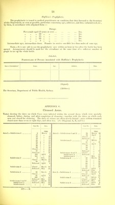
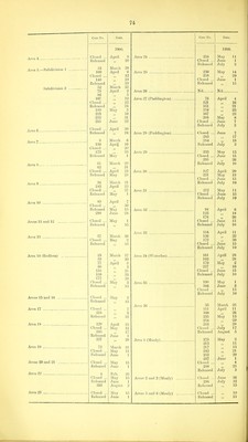
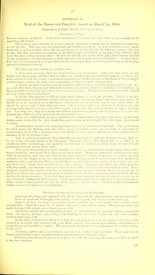
Report on the outbreak of plague at Sydney [1900-1907] / by J. Ashburton Thompson, Chief Medical Officer of the Government and President of the Board of Health.
- New South Wales. Department of Public Health
- Date:
- 1900-1908
Licence: In copyright
Credit: Report on the outbreak of plague at Sydney [1900-1907] / by J. Ashburton Thompson, Chief Medical Officer of the Government and President of the Board of Health. Source: Wellcome Collection.
Provider: This material has been provided by London School of Hygiene & Tropical Medicine Library & Archives Service. The original may be consulted at London School of Hygiene & Tropical Medicine Library & Archives Service.
84/452 page 78
![over it was slightly red, but it liad been continuously fomented witli hot boracic lotion ; there was com- mencing peri-adenitic effusion, but no matting with the smaller glands immediately above could be made out. On January llnd his temperature about midday had fallen to 99'2° F. ; he was cheerful and alert, but' pale and rather tremulous ; the headache was less ; he felt weak, but not exhausted ; the gland had markedly increased iu superficies, but was not much thickened, and though still quite tender was not at all acutely sensitive. He had slept well. On January 23rd the patient was in a similar state, but decidedly'thinner and paler than at first; his temperature had risen again to_ 102-4° F. ; the pulse was weak and easily compressed, but not intermittent. The femoral swelling had increased, and was nearly circular, about 2 inches in diameter. On January 2Uh his temperature was still lU2-4° F., and his o-eneral'state about the same ; the femoral gland vivas of the size of a mandarin orange; it was still hard, surrounded with effusion, only moderately tender, and free from fluctuation. 2.—Bacteeiological Investigation-. The bacteriology of this case was described by Dr. Frank Tidswell in the following report:— On January 21st a puncture of the gland was made with instruments just previously sterilised by exposure to steam for half an hour in the Koch steriliser. The cotton-wool wrappings were retained till the time of operation, and the instruments were still warm when used. The part had been continuously treated with boracic acid fomentations for the previous twenty-four hours. Just prior to the operation the skin was shaved, well washed with 5 per cent, carbolic lotion, then with recently boiled and still warm distilled water, and finally dried v/ith sterilised cotton wool. Through the single opening in the skin by partial withdrawal the syringe needle was passed in six different directions through tlie gland. The piston W95 raised on each occasion, but no fluid entered the barrel of the .syringe. On its final removal the channel of the needle was found to contain a minute quantity of blood. This was ejected over the surface of a serum culture tube brought for the purpose, and a second serum culture tube was inoculated from the first in the ordinary way by means of the platinum needle. Both of these tubes incubated at 37° C. remained sterile for a period of ten days, when their further observation was abandoned. On January 22nd, whilst palpating the gland, a drop of pus-like fluid was expressed through the puncture hole of the previous day. On the 23rd and 24th also similar fluid was obtained in the same way. On each occasion the single drop issuing was used to make smear preparations, and cultivations on serum and agar ; and that obtained on the 22nd was also used to inoculate a mouse. As the microscopical and cultural characters of the three samples of fluid were identical, one description will serve for all. The fluid expressed was of a dark greyish colour, showing a little rod (blood), but no yellow. A small portion set aside was found to liave clotted firmly when examined half an hour afterwards. The amount of blood present was far too small to account for this clotting, and, moreover, the clot was pale in colour. It is evident, tlierefore, that the fluid contained a large admixture of lymjih. Under the microscope the fluid was seen to contain numerous lympli cells. A few were normal in character, but most of them showed fragmented nuclei, and more or less granular cytoplasm, i.e., resembled pus cells. Lying in groups and singly amongst the cells were numerous bacilli, varying, but commonly ovoid or cylindrical in form, ranging between \fx and 3/x in length and about 'S/x broad ; ends tapering and finally rounded off; staining well with gentian violet, fuchsine, or methylene blue, and more densely at the poles than in the middle of the rods. The majority did not retain the stain when treated by Gram's method, although here and there an individual bacillus remained coloured. No spores were seen. Hounded bacterial elements were not uncommon, and the results of cultivation subsecpiently showed them to be micrococci, and not merely cocco bacilli. The cultivations were upon ordinary serum and upon nutrient agar, the inoculations being made at the bedside with the ])latinum needle sterilised in the flame of a spirit-lamp. The tubes, inoculated about noon, showed definite growth on the second morning afterwards, i.e., in about forty-five hours, having been incubated at oT^ C. in the interval. The growths were of two kinds, one composed of micrococci, the other of bacilli. The micrococcal growtli upon serum developed in the form of rounded colonies, attaining a diameter of '5 mm. iu forty-eight hours, and extending to 2 or 3 mm. in three or four days. Thick, opaque, flat, slightly irregular margins, smooth surface, creamy white in colour. In subculture the growth was more rajiid at first, a 1 mm. wide creamy streak developing in twenty-four hours. After this the growth extended slowly, reaching a width of about 3 mm. after five days at 37° G. No alteration in colour was observed. Tliere was a thick deposit and turbidity in the condensation water. U]jon nutrient agar the growth was .similar to that on serum, but whiter. On glucose agar the growth was more abundant than on nutrient agar, and was well up in twenty-four hours. In bouillon there was uniform turbidity already apparent in twenty-four hours. The cocci stained readily with gentian violet, fuchsine or methylene blue, and retained the colour when treated by Gram's method. Under tlie microscope they were seen to be regular in outline, and to lie singly or iu groups (staphylo cocci). There were no chains. Individual cocci measured 5 to '8 in diameter. The transverse line commonly seen in the pyogenic cocci was not detected in any of the specimens examined. The bacilliary growth upon serum at 37 0. was scanty. It appeared in forty-eight hours in the form of small, round, slightly raised translucent colonies, of a little less than '5 mm. in diameter. The growth had not extended much by the third day, after which, in the original tubes, it became overgrown by more rapidly developing micrococci. In subculture upon serum the growth was visible as a thin colourless streak, in forty-eight hours it increased to a band about a millimetre wide. After five days it formed a thin translucent streak still limited to the neighbourhood of the inoculation line, slightly thicker at the margins with outlying colonies, and showing granular raised specks at irregular intervals. Upon nutrient agar and glucose the growth was very simihar, and v/hen looked at from the back had a ground-glass appearance. In broth it formed abundant spicular or crumb-like particles attached to the side of the tube, with an obvious deposit of same character as that in Haffkine's prophylactic, the broth itself remaining perfectly clear and transparent. A filmy appearance on the surface disappeared on shaking, and was not reformed after four days at 37 C. In flasks with oil a film and a few pendent growths 2 to 6 mm. long were formed, as well as a copious deposit. The bacillus stained readily with violet, fuchsine, or methylene blue. Is decolourised by Gram's method, although here and there individual bacilli or small groups of them retain the colour. Non-mobile, at least as regards obvious movements of translation. The bacillus shows very distinct bipolar staining, more marked in some .specimens than in others, but clearly recognisable in all. This characteristic may amount to the colouration of only a polar granule, or to colouration of most of the bacillus, leaving only the middle of the rod unstained. Intermediate gradations are common. The bacillus varies very much in form, regularly cylindrical, boat-shaped, club-shaped, dumb-bell, and oval elements are the commonest forms. The length varies between 1 and 3 /i, and the breadth is usually about '5 fx. The ends are rounded off. No spores were observed. Some of the material obtained from the femoral swelling on 22nd January was inoculated at 4-30 p.m. into a mouse— into die back at the root of the tail. The animal was lively during all the next day. There was no visible swelling at the site of inoculation ; but the mouse was not handled. Next day it was much less lively in the morning, and got very sick during the afternoon. At 6 p.m. it was huddled up, coat rough, respiration hurried, refusint'food, but started up when the glass of its jar was flicked. It was found dead and stiff next morning at 9 a.m., having thus become definitely sick within forty-eight hours, and died within sixty-four hours after inoculation. _ The principal post-mortem features were as follows : HaBmorrhagic oedema at site of inoculation ; enlargement of the inguino-temoral glands on the right side ; no enlargement of glands detected elsewhere ; pericardium dusky, but no hemorrhages seen ; both ventricles of the heart distended with feebly clotted blood ; luncs bright red in colour, patchy pneumonia (.') ; liver definitely but not very much swollen, mottled white and pink on surface ; deep red on section ; gall bladder empty, or nearly so ; spleen not much, if at all, longer than normal, but thicker, swollen in such a way as to lose Its normal sharp edges and assume a sausage-shaped form, section deep red, trabecuh-e could not be seen with a hand lens ; stomach normal, small intestine congested, large intestines not obviously affected, but contain fluid fa;ces ; kidneys mottled, ectionpale, internal structure obscure ; bladder distended with urine of normal colour. Smear preparations and cultures made from various organs gave the bacilli showing bipolar staining. A](https://iiif.wellcomecollection.org/image/b21354704_0084.jp2/full/800%2C/0/default.jpg)