An atlas of the normal and pathological nervous systems : together with a sketch of the anatomy, pathology, and therapy of the same / Tr. and ed. (authorized) by Joseph Collins.
- Christofredo Jakob
- Date:
- 1896
Licence: Public Domain Mark
Credit: An atlas of the normal and pathological nervous systems : together with a sketch of the anatomy, pathology, and therapy of the same / Tr. and ed. (authorized) by Joseph Collins. Source: Wellcome Collection.
Provider: This material has been provided by the Augustus C. Long Health Sciences Library at Columbia University and Columbia University Libraries/Information Services, through the Medical Heritage Library. The original may be consulted at the the Augustus C. Long Health Sciences Library at Columbia University and Columbia University.
104/442 (page 40)
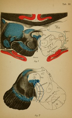
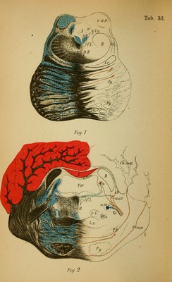
![Fig. 1. — Vertical Section through the Anterior Corpora Quad- rigemina. (Closely following that of Plate 28, 1.) The thalami (Th.o.) (pulvinar) are pressed back and outward from one another by the anterior corpora quadrigemina (e.g.a.) which press forward between them. The splenium corpus cal- losi (c.c.) with the fornix (/) lies over the anterior corpora quadrigemina. In the central gray matter (C.E.) is the aqueduct of Sylvius (A), and beneath it the nucleus of the motor oculi nerve (III) (peripheral neuron), laterally from it the nasal [descending] root of the fifth (Vn). In the corpora quadrigemina one differentiates a superficial and a deep medullary substance. Beneath these follow the tegmen- tum with the red nucleus (n.r.) of either side which gradually get closer to oue another, laterally the superior (sensory) fillet (Ls), which includes externally the inferior (of the corpora quad- rigemina) fillet {Li). Laterally to this the median geniculate body (c.g.m.). Separated from the tegmentum by the substan- tia nigra (S.n.) is the crusta (peduncle), and in its middle terri tory are the (motorial) pyramids. The optic tract (Tr.o) passes into the lateral geniculate body, the anterior [superior] quadri- geminal body, the pulvinar and there ends. From there the fibres of the optic radiation pass farther centrally (in the occipi- tal lobes). (Further details in photographic Plate 35, 2.) Fig. 2.—Section between the Anterior and Posterior Corpora Quittlrigemina. The tegmental area lias completely separated from the pulvi- nar thalami. The fillet (L) passes more caudally beneath the red nucleus. From the deep medullary substance (st.i.) the fountain-like tegmental radiation (FF) passes to the middle line where it undergoes decussation (raphe). Close beneath the motor oculi nucleus (n.lll) in the central gray matter lies the posterior longitudinal bundle which is here very clearly recog- nizable (fasciculus long, post.) (/) laterally from which the thalamus fibres pass farther (substantia reticularis). In the motor oculi nucleus are readily recognized many individual nuclei. In the posterior braohi'um (Br.a) fibres pass from the posterior quadngeminate body to the lateral geniculate body. In the substantia nigra fibres from the basal ganglion end. 40](https://iiif.wellcomecollection.org/image/b2121752x_0104.jp2/full/800%2C/0/default.jpg)