An atlas of the normal and pathological nervous systems : together with a sketch of the anatomy, pathology, and therapy of the same / Tr. and ed. (authorized) by Joseph Collins.
- Christofredo Jakob
- Date:
- 1896
Licence: Public Domain Mark
Credit: An atlas of the normal and pathological nervous systems : together with a sketch of the anatomy, pathology, and therapy of the same / Tr. and ed. (authorized) by Joseph Collins. Source: Wellcome Collection.
Provider: This material has been provided by the Augustus C. Long Health Sciences Library at Columbia University and Columbia University Libraries/Information Services, through the Medical Heritage Library. The original may be consulted at the the Augustus C. Long Health Sciences Library at Columbia University and Columbia University.
109/442 (page 41)
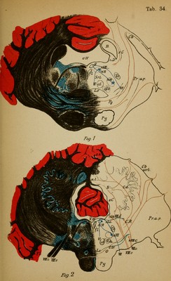
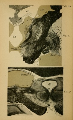
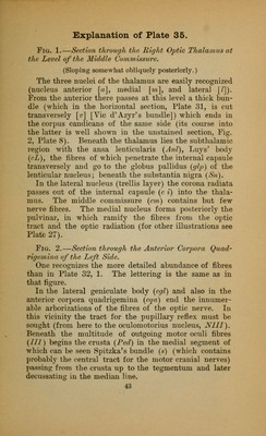
![Fig. 1.—Section through the Posterior Corpora Quadrigemina. The section includes basally the anterior portion of the pons which covers over tlie crusta. To the posterior corpora quadri- gemina (C.q.p) passes a portion of the inferior (lateral) fillet (Li). Close to the nasal (descending) trigeminus root (Vn) the trunk of the trochlear nerve which originates here is involved (IV). In the tegmentum lie the fibres of the hrachium con- junctivum, which originate in the red nucleus and decussate (D.B.) behind it (which pass to the cerebellum as the processus cerebelli ad corpora quadrigemina). At the side of the posterior longitudinal bundle (/) is the retic- ulated substance of the tegmentum. Beneath the decussation of the hrachium conjunctivum lies the horizontally placed upper (cortex-thalamus) fillet (Ls) (central sensory tract), and later- ally to it the inferior (corpora quadrigemina) fillet (Li) (cen- tral acoustic tract). One observes the enormous pontine ganglia (Pg) in which the larger part of the peduncular fibres (medial and lateral parts) end. Fig. 2.—Section through the Middle of the Pons. The aqueduct has widened into the fourth ventricle. Its roof is here formed by the velum medullare anticum (v.m.a.) with the lingula (L) (from the vermis of the cerebellum), laterally the enormous brachium conjunctivum (B) which have completed their decussation. In the tegmental territory are : the posterior longitudinal fasciculus (/), substantia reticularis tegmenti lat- eral from the raphe (R), central tegmental tract (c.t.), superior (medial) and inferior (lateral) fillet (Ls and i). Between the fibre tracts a number of cells are embedded (superior olive [01.s]. nucleus of the substantia reticularis, etc.). Laterally from the tegmentum are the motor (m) and sensory (s) nuclei of the fifth and their roots to which the nasal root of the fifth (Yn) (in its vicinity the pigmented cells of the. locus cceruleus) descends (motorial?) ; an additional tract follows from the neighboring medullary substance of the cerebellum as direct sensory cerebel- lar tract. Downward passes the caudal root of the fifth (Vc). The pyramids (Py) pass (from the peduncles) covered over by the superficial and deep pontine fibres through the pons. From the cerebellar cortex comes the processus cerebelli ad pontem (Pr.a.p.). 41](https://iiif.wellcomecollection.org/image/b2121752x_0109.jp2/full/800%2C/0/default.jpg)