An atlas of the normal and pathological nervous systems : together with a sketch of the anatomy, pathology, and therapy of the same / Tr. and ed. (authorized) by Joseph Collins.
- Christofredo Jakob
- Date:
- 1896
Licence: Public Domain Mark
Credit: An atlas of the normal and pathological nervous systems : together with a sketch of the anatomy, pathology, and therapy of the same / Tr. and ed. (authorized) by Joseph Collins. Source: Wellcome Collection.
Provider: This material has been provided by the Augustus C. Long Health Sciences Library at Columbia University and Columbia University Libraries/Information Services, through the Medical Heritage Library. The original may be consulted at the the Augustus C. Long Health Sciences Library at Columbia University and Columbia University.
53/442 (page 11)
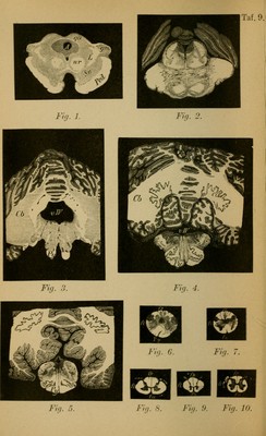
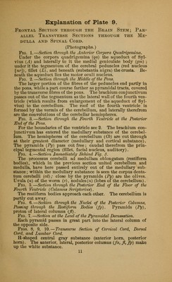
![Frontal Section through the Brain Stem; Par- allel Transverse Sections through the Me- dulla and Spinal Cord. (Photographs.) Fig. 1.—Section through the Anterior Corpora Quadrigemina. Under the corpora quadrigemina (qa) the aqueduct of Syl- vius (A) and laterally to it the medial geniculate body (gm) ; under it the tegmentum of the cerebral peduncles (red nucleus [«;■]), fillet (L), and beneath (substantia nigra) thecrusta. Be- neath the aqueduct lies the motor oculi nucleus. Fig. 2.—Section through the Middle of the Pons. The larger portion of the fibres of the peduncles end partly in the pons, while a part course farther as pyramidal tracts, covered by the transverse fibres of the pons. The brachium conjunctivum passes out of the tegmentum as the lateral wall of the fourth ven- tricle (which results from enlargement of the aqueduct of Syl- vius) to the cerebellum. The roof of the fourth ventricle is formed by the vermis of the cerebellum, and laterally therefrom are the convolutions of the cerebellar hemispheres. Fig. 3.—Section through the Fourth Ventricle at the Posterior End of the Pons. For the boundaries of the ventricle see 2. The brachium con- junctivum has entered the medullary substance of the cerebel- lum. The hemispheres of the cerebellum (Cb) are cut through at their greatest diameter (medullary and cortical substance). The pyramids (Py) pass out free; caudad therefrom the prin- cipal tegmental region (fillet, facial nucleus, auditory). Fig. 4.—Section Immediately Behind Fig. 3. The Drocessus cerebelli ad medullam oblongatam (restiform bodies), which in the previous section united cerebellum and medulla, have here passed entirely out of the medullary sub- stance ; within the medullary substance is seen the corpus denta- tum cerebelli (cd); close by the pyramids (Py) are the olives. Uvula (u) of the worm («), nodules(n) (lobes of the cerebellum). Fig. 5.—Section through the Posterior End of the Floor of the Fourth Ventricle (Calamus Scriptorius). The restiform bodies approach each other. The cerebellum is partly cut away. Fig. 6. —Section through the Nuclei of the Posterior Columns, Passing through the Restiform Bodies (fp). Pyramids (Py), proton of lateral columns (ft). Fig. 7.—Section at the Level of the Pyramidal Decussation. Each pyramid passes in great part into the lateral column of the opposite side. Figs. 8, 9, 10. — Transverse Section of Cervical Cord, Dorsal Cord, and Lumbar Cord. H-shaped central gray substance (anterior horn, posterior horn). The anterior, lateral, posterior columns (fa, fl, fp) make up the white substance. 11](https://iiif.wellcomecollection.org/image/b2121752x_0053.jp2/full/800%2C/0/default.jpg)