Results of hemisection of the spinal cord in monkeys / by Frederick W. Mott ; communicated by Professor Schäfer.
- Frederick Walker Mott
- Date:
- 1892
Licence: Public Domain Mark
Credit: Results of hemisection of the spinal cord in monkeys / by Frederick W. Mott ; communicated by Professor Schäfer. Source: Wellcome Collection.
Provider: This material has been provided by The Royal College of Surgeons of England. The original may be consulted at The Royal College of Surgeons of England.
28/70 page 26
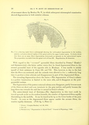
![of movement shown by Monkey No. V., in which subsequent microscopical examination showed degeneration in both anterior columns. Fig. 1. Fig. 1 is a drawing made from a photograph showing the well-marked degeneration in the median, anterior, and antero-lateral regions of the spinal cord in the upper lumbar region. It clearly shows that there are as many fibres degenerating downwards as upwards in the antero-lateral region. The preparation was made from the spinal cord of Case III. Magnification, 20 diameters. With regard to the recrossed” pyramidal fibres described by Pitres,* HoMEX,t and Sherrington;];—the latter author states that he found degenerated fibres in the crossed pyramidal tract of the opposite side in Monkeys. I was unable to satisfy myself of their existence in Cases III. and VII. This, however, is due to the fact that the fibres are scattered, and the animals were allowed to live a sufficiently long time to produce a true sclerosis and disappearance in part of the degenerated fibres. The ascending degeneration above the lesion.—The degeneration of Goll’s column in a perfect hemisection is limited to the same side, and it disappears at the post- pyramidal nucleus. The degeneration of the postero-external column soon disappears, partly because most of the fibres are short and soon terminate in the grey matter, and partly because the long fibres run towards the mid line to ascend in Goll’s column. The antero-lateral tract of Gowers.—The degeneration in this tract could be traced upwards as far as the nucleus lateralis, the number of degenerated fibres rapidly diminishing in the medulla. It can be traced as far up as the lower border of the pons Varolii. As soon as the degenerated fibres appear amidst the arcuate fibres, the number rapidly decreases. {Vide fig. 8, Plate 4.) * Pitres, ‘ Comptes Rendus,’ vol. 99, 1884. t Lor. cit. I Sherrington, “ Degeneration in Spinal Cord,” ‘ Journal of Physiology,’ 1885.](https://iiif.wellcomecollection.org/image/b22297066_0030.jp2/full/800%2C/0/default.jpg)