Results of hemisection of the spinal cord in monkeys / by Frederick W. Mott ; communicated by Professor Schäfer.
- Frederick Walker Mott
- Date:
- 1892
Licence: Public Domain Mark
Credit: Results of hemisection of the spinal cord in monkeys / by Frederick W. Mott ; communicated by Professor Schäfer. Source: Wellcome Collection.
Provider: This material has been provided by The Royal College of Surgeons of England. The original may be consulted at The Royal College of Surgeons of England.
36/70 page 34
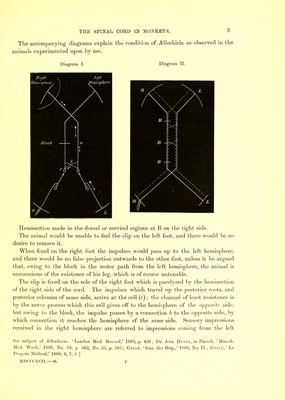
![foot. Consequently, the animal projects the sensation received from the right foot to the left foot, and it believes the cause of irritation is there, until undeceived by repeated fruitless endeavours at removal, and by visual correction. Whether this explanation be the correct one or not, it seems to me to support the view that the path of least resistance for some sensory impressions, e.g., tactile and muscular, passes up the same side of the cord, and inasmuch as the same phenomenon of Allochiria was observed in the high hemisection of the 3rd cervical segment as in the mid dorsal, upper dorsal, or lower dorsal regions, we must conclude that the main decussation does not take place till above the 3rd cervical. If we assume, as in Diagram II., that all sensory impulses coming from the foot decussate in the spinal cord, then we are unable to explain the result of this clip test. When the animals have recovered motility in the paralyzed limb, as a rule the Allochiria has passed away.* In the case where, after complete return of sensibility and almost perfect motility, the leg area was removed from the same side as the cord lesion, we saw that there was complete paralysis and greatly diminished sensibility in the previously sound leg, the animal taking no notice of the clip when placed upon the left side of the foot, but immediately removing it from the right. The disappearance of the Allochiria v'as probably due to the establishment of collateral channels other than the corpus callosum, by which sensory impressions are conveyed to the left hemisphere. Many very able experimenters—notably Munk, Schiff, Luciani, and more recently Horsley—among psychologists, Bastian and James— consider that the motor areas of the cortex cerebri are sensory, while Ferrier and others are of an opposite opinion. I cannot here enter into the arguments for or against. Mr, Horsley has told me that, after removal of portions of the cortex cerebri for disease, he has observed a sensory paralysis result on the opposite side. In connection with this I will quote Edinger,! p. 60 :— “ Sensation may also suffer from affection of the cortex of the brain. The most commonly observed symptoms are feelings of numbness, heaviness, and marked disturbance of the muscle-sense. The sense of touch is as a rule dull, so far as the judgment of the patient is concerned, but very slight sensory irritations may still be felt if they are of a simple character (such as touching with a feather, point of a needle, &c.). We do not know of any particular part of the cortex, disease of which especially leads to disturbance of sensation.” A case to the point recently occurred in Charing Cross Hospital. A young woman * [December 3, 1891.—Professor Schafer bas informed me that he has lately made a hemisection of the dorsal spinal cord in a Monkey for the purpose of studying the resulting degenex’ations. His observations entirely agree with mine as regards sensory conduction. When the animal had recovered associated movements, he removed the leg area on the opposite side ; this brought back the paralysis. Unfortunately he did not test the sensibility. I asked him to do this experiment as a complementary one to that described.] t EdingeRj “ Structure of the Central Nervous System.” Ti’anslation by Vittem and Riggs, 1890,](https://iiif.wellcomecollection.org/image/b22297066_0038.jp2/full/800%2C/0/default.jpg)