Morris' human anatomy : a complete systematic treatise / edited by C.M. Jackson.
- Sir Henry Morris, 1st Baronet
- Date:
- [1933]
Licence: Public Domain Mark
Credit: Morris' human anatomy : a complete systematic treatise / edited by C.M. Jackson. Source: Wellcome Collection.
108/1506 page 90
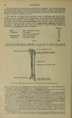
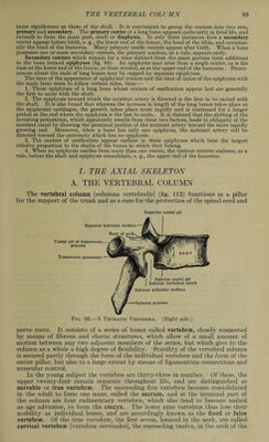
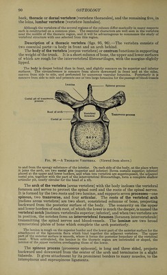
![back, thoracic or dorsal vertebrae [vertebrae thoracales], and the remaining five, in the loins, lumbar vertebrae [vertebrae lumbales]. Although the vertebra of the several regions of the column differ markedly in many respects each is constructed on a common plan. The essential characters are well seen in the vertebra near the middle of the thoracic region, and it will be advantageous to commence the study of vertebral structure with one selected from this region. Description of a thoracic vertebra (figs. 95, 96).—The vertebra consists of two essential parts—a body in front and an arch behind. The body of the vertebra [corpus vertebrae] or centrum functions in supporting the weight of the trunk. It is a short column of bone, the upper and lower surfaces of which are rough for the intervertebral fibrocartilages, with the margins slightly lipped. The body is deeper behind than in front, and slightly concave on its superior and inferior surfaces. The circumference is concave from above downward in front and at the sides, convex from side to side, and perforated by numerous vascular foramina. Posteriorly it is concave from side to side and presents one or two large foramina for the passage of blood-vessels Fig. 96.—A Thoracic Vertebra. (Viewed from above.) to and from the spongy substance of the interior. On each side of the body, at the place where it joins the arch, are two costal pits (superior and inferior) [fovea costalis superior; inferior] placed at the upper and lower borders, and when two vertebra are superimposed, the adjacent costal pits, together with the part of the intervertebral fibrocartilage, form a complete shallow articular pit, nearly circular for the head of a rib. The arch of the vertebra [arcus vertebrae] with the body incloses the vertebral foramen and serves to protect the spinal cord and the roots of the spinal nerves. It is formed by the two roots and two laminae, and supports seven processes—one spinous, two transverse, and four articular. The roots of the vertebral arch [radices arcus vertebrae] are two short, constricted columns of bone, projecting backward from the posterior surface of the body. The concavity on the upper and lower borders of each root, of which the lower is much the deeper, is named the vertebral notch [incisura vertebralis superior; inferior], and when two vertebrae are in position, the notches form an intervertebral foramen [foramen intervertebrale] transmitting the spinal nerves and blood-vessels. The laminae are two broad symmetrical plates of bone which complete the arch posteriorly. The lamina is rough on the superior border and the lower part of the anterior surface for the attachment of the ligamenta flava which bind together the adjacent vertebra. The upper part of the anterior surface is smooth, where it forms the posterior boundary of the vertebral canal. When articulated, the laminae in the thoracic region are imbricated or sloped, the laminae of the upper vertebra overlapping those of the lower. The spinous process [processus spinosus], is long and three sided, projects backward and downward from the center of the arch and terminates in a slight tubercle. It gives attachment by its prominent borders to many muscles, to the interspinous and supraspinous ligaments.](https://iiif.wellcomecollection.org/image/b31356011_0108.jp2/full/800%2C/0/default.jpg)
 No text description is available for this image
 No text description is available for this image
 No text description is available for this image