Morris' human anatomy : a complete systematic treatise / edited by C.M. Jackson.
- Sir Henry Morris, 1st Baronet
- Date:
- [1933]
Licence: Public Domain Mark
Credit: Morris' human anatomy : a complete systematic treatise / edited by C.M. Jackson. Source: Wellcome Collection.
112/1506 page 94
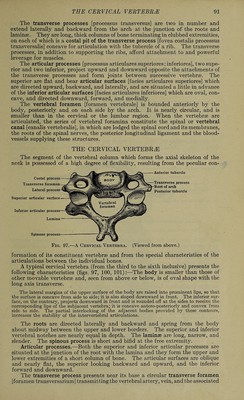
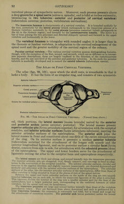
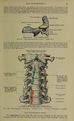
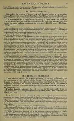
![which the atlas rotates, carrying upon it the head. It is easily recognized by the projecting odontoid process [dens] which surmounts the upper surface of the body. The anterior articular surfaces [facies articulares anteriores] are remarkable in occupying positions partly on the roots and partly on the body. They articulate with the inferior articular surfaces of the atlas. The posterior articular surfaces [facies articulares posteriores] meeting the superior articular surfaces of the third vertebra, conform to the cervical type. The odontoid process, which represents the displaced body of the atlas, is large and blunt, and bears on its anterior surface an oval saddle-form facet for articulation with the facet on the M. semispinalis M. longissimus cervicis M. iliocostalis cervicis Mm. interspinales M. trapezius M. rhomboideus minor M. serratus posterior super¬ ior M. splenius M. semispinalis capitis Transverse process of atlas M. levator scapulae (origin) M. splenius cervicis (insertion) M. levator scapulae M. splenius cervicis M. scalenus medius (insertion) M. levator scapulae M. scalenus medius M. semispinalis capitis M. levator scapulae M. scalenus medius M. semispinalis capitis and m. multifidus spinae M. rectus capitis posterior minor M. rectus capitis lateralis M. obliquus capitis superior M. obliquus capitis inferior I rectus capitis posterior major (the pointer crosses the origin of the m. obliq uus inferior . , M. semispinalis cervicis M. longissimus M. semispinalis M. longissimus M. iliocostalis cervicis M. longissimus cervicis M. iliocostalis cervicis M. semispinalis cervicis M. levator costae (origin) M. iliocostalis dorsi (inser¬ tion) Mm. interspinales M. scalenus medius M. scalenus posterior M. semispinalis and m. longissimus cap¬ itis M. multifidus M. scalenus medius M. scalenus posterior M. semispinalis and m longissimus capitis M. multifidus M. scalenus medius M. scalenus posterior M. semispinalis and m. longissimus cap¬ itis M. multifidus spinae (The large surface is for the m. multifidus) M. multifidus (and to each spinous process as high as the second) Fig. 101.—The Cervical Vertebrae. (Posterior view.) posterior surface of the anterior arch of the atlas; posteriorly it presents a smooth groove which receives the transverse ligament of the atlas. To the apex is attached the apical dental liga- ment, and to the rough surface on the side of the apex are fastened the alar ligaments which connect it with the occipital bone. The inferior surface of the body resembles that of the suc¬ ceeding vertebjge. Its anterior surface is marked by a median ridge separating two lateral depressions for the insertion of the longus colli muscle. The rfiots (pedicles), are stout and broad; the laminae are thick and prismatic: the spinous process is large and strong, deeply concave on its under surface, and markedly bifid; the trans¬ verse processes are small, not bifurcated and not grooved. The transverse foramen is directed very obliquely upward and laterally and the costal is larger than the lateral process. The anterior articular surfaces are oval, convex in the sagittal direction, and directed upward and laterally for articulation with the atlas and are like the latter in being situated in](https://iiif.wellcomecollection.org/image/b31356011_0112.jp2/full/800%2C/0/default.jpg)
 No text description is available for this image
 No text description is available for this image
 No text description is available for this image