Morris' human anatomy : a complete systematic treatise / edited by C.M. Jackson.
- Sir Henry Morris, 1st Baronet
- Date:
- [1933]
Licence: Public Domain Mark
Credit: Morris' human anatomy : a complete systematic treatise / edited by C.M. Jackson. Source: Wellcome Collection.
117/1506 page 99
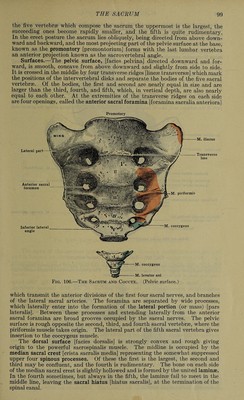
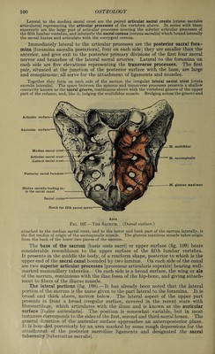
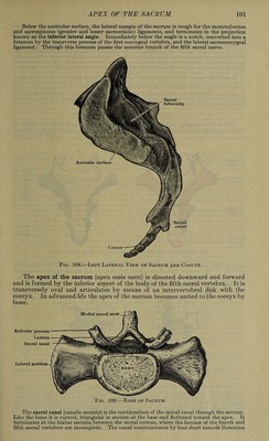
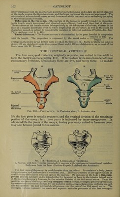
![the five vertebrae which compose the sacrum, the uppermost is the largest, the succeeding ones become rapidly smaller, and the fifth is quite rudimentary. In the erect posture the sacrum lies obliquely, being directed from above down¬ ward and backward, and the most projecting part of the pelvic surface at the base, known as the promontory [promontorium] forms with the last lumbar vertebra an anterior projection known as the sacrovertebral angle. Surfaces.—The pelvic surface, [facies pelvina] directed downward and for¬ ward, is smooth, concave from above downward and slightly from side to side. It is crossed in the middle by four transverse ridges [linese transversse] which mark the positions of the intervertebral disks and separate the bodies of the five sacral vertebrae. Of the bodies, the first and second are nearly equal in size and are larger than the third, fourth, and fifth, which, in vertical depth, are also nearly equal to each other. At the extremities of the transverse ridges on each side are four openings, called the anterior sacral foramina [foramina sacralia anteriora] Promotory Lateral part M. iliacus Transverse line Anterior sacral foramen M. piriformis Inferior lateral angle M. coccygeus M. coccygeus M. levator ani Fig. 106.—The Sacrum and Coccyx. (Pelvic surface.) which transmit the anterior divisions of the first four sacral nerves, and branches of the lateral sacral arteries. The foramina are separated by wide processes, which laterally enter into the formation of the lateral portion (or mass) [pars lateralis]. Between these processes and extending laterally from the anterior sacral foramina are broad grooves occupied by the sacral nerves. The pelvic surface is rough opposite the second, third, and fourth sacral vertebrse, where the piriformis muscle takes origin. The lateral part of the fifth sacral vertebra gives insertion to the coccygeus muscle. The dorsal surface [facies dorsalis] is strongly convex and rough giving origin to the powerful sacrospinalis muscle. The midline is occupied by the median sacral crest [crista sacralis media] representing the somewhat suppressed upper four spinous processes. Of these the first is the largest, the second and third may be confluent, and the fourth is rudimentary. The bone on each side of the median sacral crest is slightly hollowed and is formed by the united laminae. In the fourth sometimes, but always in the fifth, the laminse fail to meet in the middle line, leaving the sacral hiatus [hiatus sacralis], at the termination of the spinal canal.](https://iiif.wellcomecollection.org/image/b31356011_0117.jp2/full/800%2C/0/default.jpg)
 No text description is available for this image
 No text description is available for this image
 No text description is available for this image