Morris' human anatomy : a complete systematic treatise / edited by C.M. Jackson.
- Sir Henry Morris, 1st Baronet
- Date:
- [1933]
Licence: Public Domain Mark
Credit: Morris' human anatomy : a complete systematic treatise / edited by C.M. Jackson. Source: Wellcome Collection.
118/1506 page 100
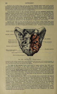
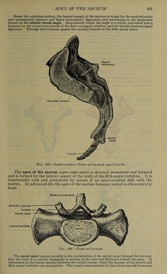
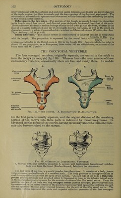
![Lateral to the median sacral crest are the paired articular sacral crests [cristse sacrales articulares] representing the articular processes of the vertebrae above. In series with them superiorly are the large pair of articular processes meeting the inferior articular processes of the fifth lumbar vertebra, and inferiorly the sacral cornua [cornua sacralia] which bound laterally the sacral hiatus and articulate with the coccygeal cornua. Immediately lateral to the articular processes are the posterior sacral fora¬ mina [foramina sacralia posteriora], four on each side; they are smaller than the anterior, and give exit to the posterior primary divisions of the first four sacral nerves and branches of the lateral sacral arteries. Lateral to the foramina on each side are five elevations representing the transverse processes. The first pair, situated at the junction of the posterior surface with the base, are large and conspicuous; all serve for the attachment of ligaments and muscles. Together they form on each side of the sacrum the irregular lateral sacral crest [crista sacralis lateralis]. The space between the spinous and transverse processes presents a shallow concavity known as the sacral groove, continuous above with the vertebral groove of the upper part of the column, and, like it, lodging the multifidus muscle. Bridging across the groove and Articular Auricular Median sacral crest Articular sacral crest [Lateral sacral crest Posterior sacral foramen M. multifidus M. sacrospinalis M. gluteus maximus Hiatus sacralis leading in¬ to the sacral canal Sacral cornu Notch for fifth sacral Apex Fig. 107.-—The Sacrum. (Dorsal surface.) attached to the median sacral crest, and to the lower and back part of the sacrum laterally, is the flat tendon of origin of the sacrospinalis muscle. The gluteus maximus muscle takes origin from the back of the lower two pieces of the sacrum. The base of the sacrum [basis ossis sacri] or upper surface (fig. 109) bears considerable resemblance to the upper surface of the fifth lumbar vertebra. It presents in the middle the body, of a reniform shape, posterior to which is the upper end of the sacral canal bounded by two laminae. On each side of the canal are two superior articular processes [processus articularis superior] bearing well- marked mammillary tubercles. On each side is a broad surface, the wing or ala of the sacrum, continuous with the iliac fossa of the hip-bone, and giving attach¬ ment to fibers of the iliacus muscle. The lateral portions (fig. 108).—It has already been noted that the lateral portion of the sacrum is the name given to the part lateral to the foramina. It is broad and thick above, narrow below. The lateral aspect of the upper part presents in front a broad irregular surface, covered in the recent state with fibrocartilage, which articulates with the ilium and is known as the auricular surface [fa.cies auricularis]. The position is somewhat variable, but in most instances corresponds to the sides of the first, second and third sacral bones. The general direction of the auricular surface approaches an anteroposterior plane. It is bounded posteriorly by an area marked by some rough depressions for the attachment of the posterior sacroiliac ligaments and designated the sacral tuberosity [tuberositas sacralis].](https://iiif.wellcomecollection.org/image/b31356011_0118.jp2/full/800%2C/0/default.jpg)
 No text description is available for this image
 No text description is available for this image
 No text description is available for this image