Morris' human anatomy : a complete systematic treatise / edited by C.M. Jackson.
- Sir Henry Morris, 1st Baronet
- Date:
- [1933]
Licence: Public Domain Mark
Credit: Morris' human anatomy : a complete systematic treatise / edited by C.M. Jackson. Source: Wellcome Collection.
120/1506 page 102
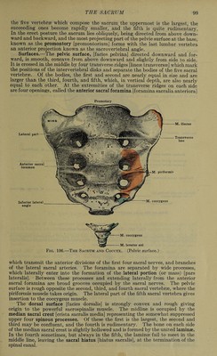
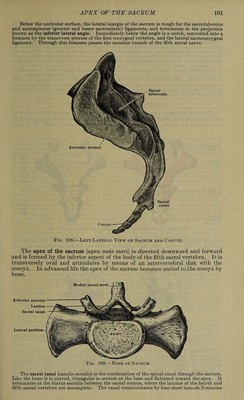
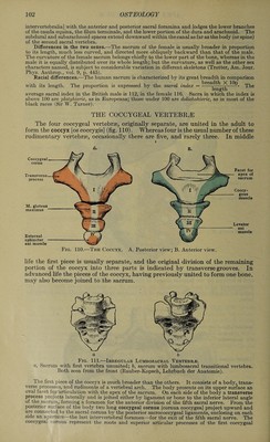
![intervertebralia] with the anterior and posterior sacral foramina and lodges the lower branches of the cauda equina, the filum terniinale, and the lower portion of the dura and arachnoid. The subdural and subarachnoid spaces extend downward within the canal as far as the body (or spine) of the second sacral vertebra. Differences in the two sexes.—The sacrum of the female is usually broader in proportion to its length, much less curved, and directed more obliquely backward than that of the male. The curvature of the female sacrum belongs chiefly to the lower part of the bone, whereas in the male it is equally distributed over its whole length; but the curvature, as well as the other sex characters named, is subject to considerable variation in different skeletons (Trotter, Am. Jour. Phys. Anthrop., vol. 9, p. 445). Racial differences.—The human sacrum is characterized by its great breadth in comparison with its length. The proportion is expressed by the sacral index = -*^ ^• The average sacral index in the British male is 112, in the female 116. Sacra in which the index is above 100 are platyhieric, as in Europeans; those under 100 are dolichohieric, as in most of the black races (Sir W. Turner). THE COCCYGEAL VERTEBRAE The four coccygeal vertebrae, originally separate, are united in the adult to form the coccyx [oscoccygis] (fig. 110). Whereas four is the usual number of these rudimentary vertebrae, occasionally there are five, and rarely three. In middle A. M. gluteus maximus External sphincter ani muscle Coccygeal cornu process Fig. 110.—The Coccyx. A. B. Facet for apex of sacrum Coccy- geus muscle Levator ani muscle Posterior view; B. Anterior view. life the first piece is usually separate, and the original division of the remaining portion of the coccyx into three parts is indicated by transverse grooves. In advanced life the pieces of the coccyx, having previously united to form one bone, may also become joined to the sacrum. a b Fig. 111.—Irregular Lumbosacral Vertebra. a, Sacrum with first vertebra ununited; b, sacrum with lumbosacral transitional vertebra. Both seen from the front (Rauber-Kopsch, Lehrbuch der Anatomie). The first piece of the coccyx is much broader than the others. It consists of a body,, trans¬ verse processes, and rudiments of a vertebral arch. The body presents on its upper surface an oval facet for articulation with the apex of the sacrum. On each side of the body a transverse P£0('ess projects laterally and is joined either by ligament or bone to the inferior lateral angle of the sacrum, forming a foramen for the anterior division of the fifth sacral nerve. From the posterior surface of the body two long coccygeal cornua [cornua coccygea] project upward and are connected to the sacral cornua by the posterior sacrococcygeal ligaments, enclosing on each side an aperture the last intervertebral foramen—for the exit of the fifth sacral nerve. The coccygeai cornua represent the roots and superior articular processes, of the first coccygeal](https://iiif.wellcomecollection.org/image/b31356011_0120.jp2/full/800%2C/0/default.jpg)
 No text description is available for this image
 No text description is available for this image
 No text description is available for this image