Morris' human anatomy : a complete systematic treatise / edited by C.M. Jackson.
- Sir Henry Morris, 1st Baronet
- Date:
- [1933]
Licence: Public Domain Mark
Credit: Morris' human anatomy : a complete systematic treatise / edited by C.M. Jackson. Source: Wellcome Collection.
84/1506 page 66
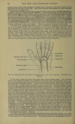
![vening furrows [sulci cutis] arranged in parallel lines in groups on the flexor surface of the hands and feet are also of this type. They form patterns characteristic for each individual and permanent throughout life. Among the projections are the large permanent folds of skin such as the labia pudendi, the preputium penis, the frenula preputii clitoridis, and labiorum pudendi, and less marked ridges as the median raphe of the perineum, scrotum and penis, and the tuberculum labii superioris. Of a somewhat different sort are the touch pads [toruli tactiles] of the hands and feet. Among the larger depressions in addition to the umbilical fovea, is the coccygeal foveola, and a consider¬ able number of well-marked permanent furrows found in various places, such as the nasolabial and mentolabial sulci, the philtrum labii superioris, the infraorbital sulcus, and the infraorbital and supraorbital palpebral sulci. There are numerous articular furrows on both the flexor and extensor surfaces produced by the action of the joints, and associated with intervening folds of skin, particularly on the dorsal surface. They are especially noticeable on the hands. Variations of the palmar joint-sulci are due to variations in opposition of the thumb and the use of the fingers and the relative arrangement of the thumb and fingers and joints. They are of importance as indicating topographically the position of the joints, their relation to which has been made clearer by means of the X-ray. Two or three of these are seen on the palmar surface of the wrist; two lower down and usually close together, and one less well marked, a little higher up upon the forearm (fig. 74). None of these corresponds exactly to the wrist-joint. The lowest ‘precisely crosses the, arch of the os magnum in the line of the third metacarpal bone’ (Tillaux), and is not quite 1.8 cm. (% in.) below the arch of the wrist- joint. It is about 1.2 cm. in.) above the carpometacarpal joint line, and indicates very fairly the upper border of the transverse carpal (anterior annular) ligament. •-Inferior fold •••Middle fold — Superior fold - —Ulnar artery Fig. 71.—Relation of the Folds of the Palm to the Volar Arches. (Modified from Tillaux.) Of the many creases in the skin of the palm, three require especial notice. The first starts at the wrist, between the thenar and hypothenar eminences, and, marking off the former emi¬ nence from the palm, ends at the lateral border of the hand and at the base of the index-finger. The second fold is slightly marked. It starts from the lateral border of the hand, where the first fold ends. It runs obliquely medially across the palm, with a marked inclination toward the wrist, and ends at the lateral limit of the hypothenar eminence. The third, lowest, and best marked of the folds starts from the little elevation opposite the cleft between the index and middle fingers, and runs nearly transversely to the ulnar border of the hand, crossing the hypo¬ thenar eminence at the upper end of its lower fourth. The first fold is produced by the adduc¬ tion of the thumb; the second, mainly by the bending simultaneously of the metacarpophalan¬ geal joints of the first and second fingers; and the third by the flexion of the three medial fingers. The second fold, as it crosses the third metacarpal bone, about corresponds to the lowest part of the superficial volar arch. The third fold crosses the necks of the metacarpal bones, and indicates pretty nearly the upper limits of the synovial sheaths for the flexor tendons of the three lateral fingers. A little way below this fold, the palmar aponeurosis breaks up into its four slips, and midway between the fold and the webs of the fingers lie the metacarpophalan¬ geal joints. Of the transverse folds across the fronts of the fingers, corresponding to the meta¬ carpophalangeal and interphalangeal joints, the highest is placed nearly 18 mm. in.) below its corresponding joint. The middle folds are multiple for all the fingers, and are exactly opposite to the first interphalangeal joints. The distal creases are single, and are placed a little above the corresponding joints. There are two single creases on the thumb corresponding to the two joints, the higher crossing the metacarpophalangeal joint obliquely. The free edge of the web of the fingers, measured from the palmar surface, is about 1.8 cm. (,% in.) from the metacarpophalangeal joints. (Treves.) The folds and furrows brought about through the action of the skin muscles run at right angles to the muscle fibers and are more or less transitory at first but become more permanent through repeated or long-continued action. They are represented by the wrinkles of the fore-](https://iiif.wellcomecollection.org/image/b31356011_0084.jp2/full/800%2C/0/default.jpg)
 No text description is available for this image
 No text description is available for this image
 No text description is available for this image