Nervous and mental diseases / by Archibald Church and Frederick Peterson. With 338 illustrations.
- Church, Archibald, 1861-1952
- Date:
- 1903
Licence: Public Domain Mark
Credit: Nervous and mental diseases / by Archibald Church and Frederick Peterson. With 338 illustrations. Source: Wellcome Collection.
Provider: This material has been provided by the Augustus C. Long Health Sciences Library at Columbia University and Columbia University Libraries/Information Services, through the Medical Heritage Library. The original may be consulted at the the Augustus C. Long Health Sciences Library at Columbia University and Columbia University.
73/956 page 71
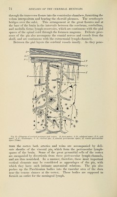
![PAET II. DISEASES OF THE CEREBRAL MENINGES AND CRANIAL NERVES. CHAPTER I. THE CEREBRAL MENINGES—PACHYMENINGITIS AND PIAL HEMORRHAGE. Anatomical Considerations.—The coverings of the brain are admirably suited to protect it from injury and infection. Guarded ex- ternally by the skull and the scalp-pad, it is intimately enveloped by the dense, fibrous dura mater in a practically sealed sac. The usual anatom- ical descriptions of the cerebral meninges are misleading. Ordinarily three distinct membranes are named and described, when in reality there are but two. Lining the skull we have the dura mater, serving as an internal periosteum for the cranial bones and furnishing in ])art their vascular supply. It is entirely free from the brain, but gives off sheaths to the cranial nerves and the large vessels at their exit from the skull, and supplies venous channels or sinuses for the return circulation of the encephalon. The dural fold between the cerebral hemispheres and the tentorium cerebelli afford support and protection. In normal conditions the brain fills the cranial cavity fully, and its soft covering membrane is everywhere in contact with the inner surface of the dura. The interval which separates them is called the subdural space. No actual space, however, exists, the two membranes being smoothly applied to each other and only separated by disease or mechan- ical means. Closely investing the brain is the jikt mater, made up of two layers or membranes very loosely attaclied by delicate meshes of fibrous tissue. The outer can be easily stripped from the under layer, and constitutes what is usually described as the arachnoid. The alleged double layers and spaces of the so-called arachnoid can not be demonstrated and do not exist. This outer pial layer we may call the arachnopia. Between it and the under layer, or visceralpia, is a varying space, the p>kd space, filled with a delicate, open, reticular network of fibrous tissue containing cerebrospinal fluid or lymph. It is an enormous lymph-space. At the gyral grooves the visceral pia dips to the bottom of the sulci. It everywhere closely adheres to the brain-cortex, which it follows](https://iiif.wellcomecollection.org/image/b21225953_0073.jp2/full/800%2C/0/default.jpg)