Content advisory
This digitised material is free to access, but contains information or visuals that may:
- include personal details of living individuals
- be upsetting or distressing
- be explicit or graphic
- include objects and images of objects decontextualised in a way that is offensive to the originating culture.
Licence: In copyright
Credit: "Nucleic acids". Source: Wellcome Collection.
40/64
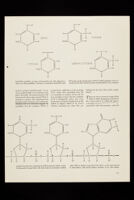
![MODEL OF DNA STRUCTURE is a double helix with slanted cross-links connecting the two chains. The nucleic acid chains which form the helixes are identical but head in opposite direc- ss-links (black lines) consist of complementary pairs connected by hydrogen bonds. The broken colored 3 centers of the cross-links and also forms a helix. with any other part of the cell. Each set of chromosomes appears to have a fixed amount of DNA. There are at least two phenomena which give direct evidence that DNA plays a genetic role. Firstly, it has been found that pure DNA extracted from certain bacteria is capable of trans ferring some of the properties of this strain to a related strain, and the trans formed bacteria pass these properties on to their descendants. Secondly, when a bacterial virus infects a bacterium, it is the virus's DNA (not its protein) that enters the bacterial cell, and much of this DNA turns up in the progeny virus produced in the cell. Taking all the evidence together, it is difficult to resist the conclusion that DNA is genetic material. If that is the case, our problem is to learn how DNA reproduces itself. The double-helical structure of DNA suggests a possible answer, which I have discussed in a previous article [see The Structure of the Hereditary Material, by F. H. C. Crick; Scientific American , October, 1954]. The basic idea is that the two RADIOACTIVITY OF LABELED DNA in a bacterial virus is indicated by this star of tracks in a nuclear-emulsion photograph made by Cyrus Levinthal of the Massachusetts Institute of Technol ogy. When bacteria infected with the virus were grown in a medium containing radioactive phosphorus, the viruses used the radioactive material in making new DNA. Then viruses containing radioactive DNA were transferred to a culture of bacteria growing in a non radioactive medium. When the bacteria were broken open by the virus infection, their contents were mixed with the photographic emulsion. Each track in the developed emulsion is made by a par ticle emitted in the decay of a phosphorus atom; the number of tracks is proportional to the amount of radioactive phosphorus in the virus. Thus it is possible to trace how the phosphorus in the DNA of first generation of viruses is distributed in later generations.](https://iiif.wellcomecollection.org/image/b18172957_PP_CRI_I_1_14_0040.jp2/full/800%2C/0/default.jpg)




 No text description is available for this image
 No text description is available for this image
 No text description is available for this image