An introduction to pathology and morbid anatomy / by T. Henry Green.
- Green, T. Henry (Thomas Henry), 1841-1923
- Date:
- 1900
Licence: In copyright
Credit: An introduction to pathology and morbid anatomy / by T. Henry Green. Source: Wellcome Collection.
Provider: This material has been provided by The University of Leeds Library. The original may be consulted at The University of Leeds Library.
70/618 (page 54)
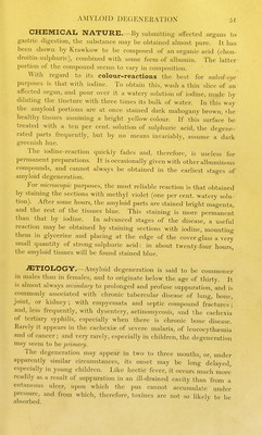
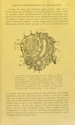
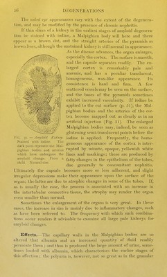
![To the linked cijc, the amyloid liver possesses the typical cha- racters already descri])ed (p. 50). If the change is very far advanced, the tissue may be perfectly homogeneous, all distinction between the individual lobules being lost. In other cases the lobules are distinctly mapped out; they are enlarged, and the external zone may be of an opaque yellowish-white colour owing to the presence of fat. This association of the fatty and amyloid changes is exceedingly common. Amyloid degeneration does not obstruct the portal circu- lation, and hence does not cause ascites (see Cirrhosis of Liver), except in those rare cases in which the portal vessels are involved. It causes fatty degeneration and atrophy of the hepatic cells, and thus interferes with the functions of the or<ran. o If sections are stained with iodine, the mahogany colour will frequently be found limited to the so-called intermediate zone of the lobules—the area of distribution of the hejiatic artery. The appearance thus produced is that of a number of partially com23ressed rings with pale centres, and still paler intervening spaces (Fig. 29). Thus the earliest seat of amyloid degeneration differs from that of fatty infiltration, in which the fat first accumulates in the cells of the outer or 2)ortal zone (Fig. 15), and from that of passive congestion, in which the changes beg-in in the central zone around the intralobular vein. All these changes not imcommonly occur together. As the amyloid change advances, first the central zone and later on the peripheral zone are affected, and even the interlobular connective-tissue may ultimately become involved. Amyloid Degeneration of the Kidneys. Microscopical 11/, the degeneration is first observed in the Malpighian bodies (Fig. 30). At first only a few of the capillary loops in each tuft are affected, but all tlie loops gradually become involved. The whoU: coil then presents an ill-defined outline and glistening surface. The cliange in tlie meantime extends to the afferent arteries, to the c.ipillary network around the tubules, to the arteriole rectae of the medulla, and, in advanced cases, to the intertubular tissue and to the tunica propri.n of the tubules. It is doubtful if the epithelium ever undergoes amyloid degeneration. The distribution of the change may be very irregidar. Fig. 29.—Amyloid Liver. Stained with iodine. The darkest portions represent the affected intermediate zones. Natural size.](https://iiif.wellcomecollection.org/image/b21503060_0070.jp2/full/800%2C/0/default.jpg)