Licence: Public Domain Mark
Credit: Ophthalmic surgery / by J. F. Streatfield. Source: Wellcome Collection.
Provider: This material has been provided by UCL Library Services. The original may be consulted at UCL (University College London)
49/56 page 47
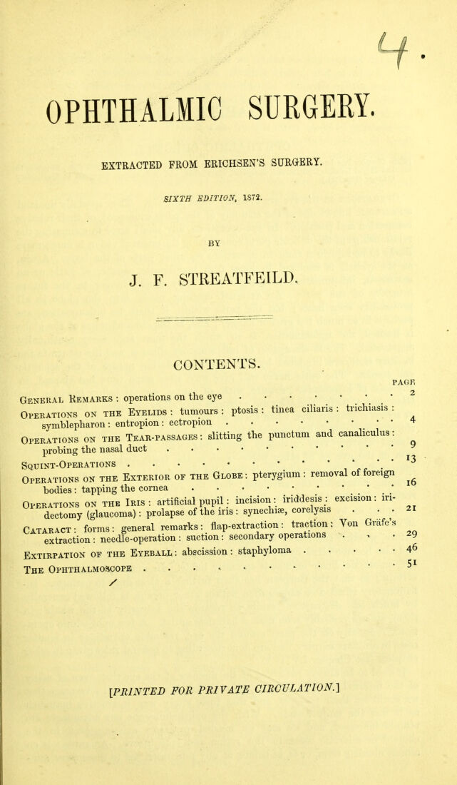
![EXTIllFATION OF THE EYEBALL. l>ein;^' placed opposite to a noiseloss gas-burner, or other very bright light, but not so near that the heat of the lianie is ai)pret iable ; ^vhen (juestioned very many tinifs, tilt' patient, if blind, cannot rightly tell the Hanie from the absence of it, when it is turned down so low that no actual light remains. The patients have had tlie power of vision, they may be habitually truthful, but they are glad to de- ceive themselves unconsciously (we need not tell them so); they feel the warmth of the sun's rays, or of the fire, and, knowing what is the cause of this, they think tliat, as they used, they still perceive the accompanying liglit : they hear the shutters shut and the lanq) set on the table, and they picture to themstdves the light, and even the objects lit up l)y it, in their accustomed positions. Or, they subjectitrlij see Hashes of light, which are symptomatic of their complaint, and are analogous to the liglit we see in the dark with our eyes shut when the eyelids are violently rubbed. They say they can fmd their way about, but tlien it is in their own homes. Tliey say they can count fingers, and then they hold up their own to count. They are loth to believe that they really see notliing at all, and therefore it is that in all cases it is necessary rigidly to cross-examine every patient in whose case we have reason to suspect total blindness or the absence of any perception of light. A few questions will be only misleading, and every such case must be treated as if we suspected the l)atient of malinfjerituj, as it is Cidled by army surgeons. Thk Operation is thus done. When the i)atient is under the influence of an amesthetic, a (spring) wire speculum is inserted between the lids to keep them open ; the ccjnjunctiva is seized with toothed forceps, at any part near the margin of the cornea, ami a small opening is made in it with rather blunt-pointed scissors. In the next place, the conjunctiva is to be divided all around the cornea. This may be done by inserting one blade of the scissors beneath the conjunctiva where it has been opened, and drawing the globe with the forceps one way or the other, as may be most convenient; or by seizing the conjunctiva with the forceps at the i)art to be next cut through, if it be too closely adherent to the subjacent parts to allow tlie passage of the point of the scissors. Or the stra- bismus-hook (Fig. 453, (/), inserted at the opening made in the conjunctiva, may be carried round the cornea, and the conjunctiva, raised a little in this w^ay, may be conveniently cut through if it be loose. When the circular incision of the mucous membrane is completed, the subconjunctival tissues, seized at dilferent ]>arts in the wound, are cut through with the scissors, the points being directed backwards and kept as closely as i)ossil)le to the globe. An open- ing blill deeper, close to the globe, in some part of the circumference of the wound, ]> made with the scissors, and then the strabismus-hook is passed beneath the tendon of one or other of the rectus muscles, which is divided, and so are then the otlier tendons in succession and otlier intervening parts close to and connected ^\■ith the globe. ^ The tendons of the obli(pie muscles being directed to the tem- l>oral side to be inserted, the hook must Ije directed outwards and kept very close to the globe to secure them. But only the four rectus muscles are generally w uvih^ a separate search. All the tendons being cut through, the divided ocular insertion of the internal rectus is seized with the forceps and the eye drawn out- wards ; a pair of scissors curved on the flat (Fig. 453, e) is carried down beside the globe on the nasal side, as, on this side, the optic nerve enters the globe and is most easily reached ; the scissors are opened a little and advanced, and the nerve is at one stroke divided. The globe now generally comes forward suddenly. It is drawn forward by holding it at the point of insertion of one of the rectus muscles or wherever it may be secured conveniently, and any remaining adherent soft parts are divided, always of course as before, close to the globe. Dixon finds it convenient to leave the insertion of the](https://iiif.wellcomecollection.org/image/b21636953_0051.jp2/full/800%2C/0/default.jpg)



 No text description is available for this image
 No text description is available for this image
 No text description is available for this image