Text-book of skin diseases / by Dr. Isidor Neumann ... ; translated from the 2nd German edition, by special permission of the author, by Alfred Pullar.
- Neumann, Isidor von, 1832-1906.
- Date:
- 1871
Licence: Public Domain Mark
Credit: Text-book of skin diseases / by Dr. Isidor Neumann ... ; translated from the 2nd German edition, by special permission of the author, by Alfred Pullar. Source: Wellcome Collection.
Provider: This material has been provided by University of Bristol Library. The original may be consulted at University of Bristol Library.
329/360 page 305
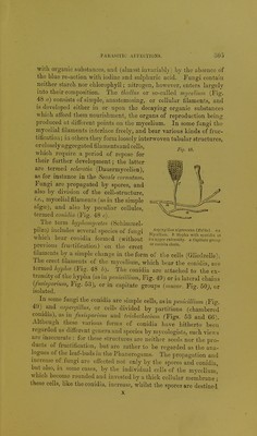
![Fig. 48. with organic substances, and (almost invariably) by the absence of the blue re-action witli iodine and sulphuric acid. Fungi contain neither starch nor chlorojDhyll; nitrogen, hovrever, enters largely into their composition. Tlie thallus or so-called mycelium (Fig. 48 a) consists of simple, anastomosing, or cellular filaments, and is developed either in or upon the decaying organic substances which afibrd them nourishment, the organs of reproduction being produced at dijSferent points on the mycelium. In some fungi the mycelial filaments interlace freely, and bear various kinds of fruc- tification ; in others they form loosely interwoven tubular structures, or closely aggregated filaments and cells, which require a period of repose for their further development; the latter are termed sclerotia (Dauermycelien), as for instance in the Secale cornutum. Fungi are propagated by spores, and also by division of the cell-structure, i.e., mycelial filaments (as in the simple algoo), and also by peculiar cellules, termed conidia (Fig. 48 c). The term hyphomycetes (Schimmel- pilza; includes several species of fungi which bear conidia formed (without previous fructification) on the erect filaments by a simple change in the form of the cells (Gliedzelle). The erect filaments of the mycelium, which bear the conidia, d]'o termed hyphce. (Fig. 48 h). The conidia are attached to the ex- tremity of the hypha (as in penicillium, Fig. 49) or iu lateral chains (fusisporium, Fig. 53), or in capitate groups [mucor. Fio-. 50), or isolated. ° In some fungi the conidia are simple cells, as in penicillium (Fig. 49)^ and aspercjillus, or cells divided by partitions (chambered conidia), as in fusisporium and triclioihecium (Figs. 63 and 66). Although these various forms of conidia have hitherto been regarded as different genera and species by mycologist s, such views are inaccurate : for these structures are neither seeds nor the pro- ducts of fructification, but are rather to be regarded as the ana- logues of the leaf-buds in the Phanerogams. The propagation and increase of fungi arc effected not only by the spores and conidia, but also, in some cases, by the individual cells of the mycelium, which become rounded and invested by a thick cellular membrane • these cells, like the conidia, increase, whilst the spores are destined Asprr^'illus nigrescens (Holin). an. My>:eliam. b. Hypha with sporidi.i at its upper extremity, c. Capitate group or conidia chain.](https://iiif.wellcomecollection.org/image/b21445904_0329.jp2/full/800%2C/0/default.jpg)