Atlas and abstract of the diseases of the larynx / by L. Grünwald ; authorized translation from the German edited by Charles P. Grayson.
- Ludwig Grünwald
- Date:
- 1902
Licence: Public Domain Mark
Credit: Atlas and abstract of the diseases of the larynx / by L. Grünwald ; authorized translation from the German edited by Charles P. Grayson. Source: Wellcome Collection.
Provider: This material has been provided by the Augustus C. Long Health Sciences Library at Columbia University and Columbia University Libraries/Information Services, through the Medical Heritage Library. The original may be consulted at the the Augustus C. Long Health Sciences Library at Columbia University and Columbia University.
26/284 page 20
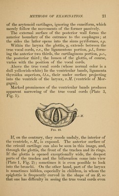
![of the aperture of the larynx, the anterior parts appear above, and the posterior parts below. It is only when the image is represented on paper that it appears com- pletely inverted, the front corresponding to the back, and vice verna. U.s c.v. Fig. 9 shows the laryngoscopic image of a normal larynx as it appears on paper, the movable parts in the position which they occupy during respiration. It also shows the anatomy of the parts in the living subject. E., epiglottis ; in the middle is seen its posterior surface, which is rolled from behind forward and therefore looks uj)ward ; on each side the surface of the tongue is covered witli blood-vessels. This is always taken as tlie starting- ])()int in laryngoscopy, the directions being given accord- ing to the actual relations of the parts, not the one that apj)ears in the image. In front of the epiglottis the val- lecula (V.) extends to the tongue, Z., interrupted at its center by the glosso-e])iglottidean ligament, Lg.e. The posterior margin of the cavity of the larynx begins at the sides witli tlie aryteno-epiglottidean folds, Ld.c, wliich invest the cartilages of Wrisberg and Santo- rini. The arytenoid cartilages lie hidden beneath them, and between these cartilages tlie mucous membrane dips down into the interarytcnoid sj)ace. For the sake of brevity in describing the relations of parts, we speak only](https://iiif.wellcomecollection.org/image/b21220463_0026.jp2/full/800%2C/0/default.jpg)


 No text description is available for this image
 No text description is available for this image
 No text description is available for this image