The anatomy of humane bodies, with figures drawn ... by some of the best masters in Europe ... To which is added an introduction explaining the animal oeconomy / Revised and publish'd by C.B. Albinus.
- William Cowper
- Date:
- 1737
Licence: Public Domain Mark
Credit: The anatomy of humane bodies, with figures drawn ... by some of the best masters in Europe ... To which is added an introduction explaining the animal oeconomy / Revised and publish'd by C.B. Albinus. Source: Wellcome Collection.
55/380
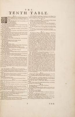
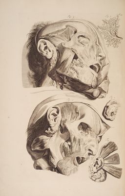
![TENTH Fig. 1. Verse HE Brain eres with the Medulla Oblongata continu’d sw to it, when free’d from the Skull, and Specus or Cavity of all the Vertebre of the Neck, Back, and Loins. 3 ¥ AA, The Dura Mater free’d from the Brain, and yy WANG fome what expanded. prema t 2a, Part of the Falx. BB, Part of the Brain cut Tranfverfly. — C, The Divifion in its Cortical Part, which compofe thofe turnings, and windings on its External Surface. DD, The Cortical, or Cinericious Part of the Brain; by fome call’d the Glandulous Part: EE, The Medullary, or white Part of the Brain; by fome call’dthe Callous, and Fibrous Part. ) ff, The Hindmoft Part of the Brain, which refted on the Second Procefs of the Dura Mater. FGH, The Right and Left Ventricles of the Brain open’d; where the Blood Vefiels of the Pia Mater, which Line them, may be feen: F, their Upper and Foreparts, which are largeft, and become ftill lef, and lefs towards their Lower, and Back-parts, G. HA, The Corpus Callofum. IK, The Roots of the Fornix. L, The Zhalamus Nervi Optici of the Right Side ; that of the Left, not being Letter’d. M, The Corpus Tranfverfale of the Corpus Callofum. NN, Parts of the Corpora Striata whole. OO, The Nates. PP, The Tefes. Q, The Glandula Pinealis, in fitu. RR, The Plexus Coroeides compos’d of Blood Veflels of both Kinds, Lympheducts, Membranes, and Glands. See Fig. 3. SS, The Firft Procefs of the Cerebellum, going to the Nates. T, A Tranfverfe Procefs joyning the Two Pathetick Nerves , and laft mentioned Procefs, f{, The Fourth Ventricle, call’d Calamus Scriptorius. VV, The Pathetick Nerves, W W , Two Procetles of the Spinal Marrow which compofe the Sides of the Fourth Ventricle. XYZ, The Meditullium of the Cerebellum appearing in an Arboreous Manner, after a Tranfverfe Scétion of the Cerebellum. ab, ab, €fc.'The Dura Mater , which inclofes the Spinal Marrow, divided, and expanded. cc, &c. The Pia Mater as yet inclofing the ‘Medulla Spinalis, but raifed witha Probe in its Lower Part, where itinverts the Cauda Equina. 123,€9¢. The feveral Pairs of Nerves {pringing from the Medulla Spinalis: From 1 to g the Origins of the Nerves of the Neck ; the Firft of which pafles out at the Third Perforation of the Os Occipitis , and ts reckoned the Tenth of the Brain; the reft march out between the Vertebre of the Neck, Back, Loins, and Perforations of the Os Sacrum fucceflively ; that of Fig. 9 marching out between the Sixth and Seventh Vertebra of the Neck; thofe of 10 to 21 are the Nerves of the Back: From 22 to 27 thofe of the Loins; the reft go out at the Foramina of the Os Sacrum. : Fig. 2. AA, Part of the Brain bosf'd, and view’d with a Microfcope. BB, The Membranes of the Brain {eparated ; of which the External 1s the Dura Mater; the Two Internal compofe the Pia Mater. CD, The Reticular Diftribution of the Blood Veffels near their Ex- tremities. EE, Divers Orders of Cortical Glands on the Surface of the Brain. FF, The Tubes deriv’d from thofé Cortical Glands. GG, The Lobes, or diftin& Clufters of Glands wreathed with va- rious Angles. HH, The Complicated Tubes. . II, The Nervous Fibres deriv’d from the laft mentioned Tubes. Yi arg ‘ of the Plexus Coroeides dlinestes , by the help of a Magnifying afs, AA, The Membranous inclofures of the Fa/ciculi of the Veflels, feparated. BC, The Blood Veffels extended with Plaifter of Paris, and their own Blood. | | DD, Branches of Lympheduéts, fomewhat extended with Wind. E, Nervous Tubuli according to Bidloo, which I can by no means conceive to be exiftent in the Plexus Coroeides. FF, The Glands of the Plexus Coroeides placed irregular, of which, fome are Hard, and Fibrous, others are Veliculous, and Flaccid. Fi An _ A Portion of the Medulla Oblongata cut off, and divided laterally ac- cording to its Length; expreft fomwhat bigger than the Life. _ AA, The Upper Part of the Atedulla Oblongata. BB, The Fore and Back Part. CC, The Nervous Fibrille arifing from the Fore, and Back Partof the Spinal Marrow. DD, The Inferior Part of the Spinal Marrow cut off. EE, Portions of the Dura Mater left, to fhew its Perforations for the Netves , as they pafs out of the Specus of the Vertebre, TABLE. FFF, The Plexus Ganglioformes of the Nerves at their Egrefs from between the Vertebre: Two or Three of the Bodies of the Nervesthem- {elyes are expreft in this Figure pinn’d out. Fig. 5. 7 | A Portion of the Medulla Spinalis, cut off about the Third Pertebra of the Back, expreft fomewhat bigger than the Life. A, The Upper Part of the Spinal Marrow. BB, A Portion of the Continuation of the Dura Mater expanded. | CC, The Nervous Fibres arifing from the Fore and Back Parts of the Spinal Marrow. . D, The Nervous Fibrille collectively pafling thro? the Dura Mater. E, Their Gangliform Plexus at the Beginnings of the Bodies of the Nerves. FE, A Divifion of the Spinal Marrow according to its Length. G, Some Veftigia of Blood Veflels, which pais on the Outfide of the Spinal Marrow, Fig. 6 The Structure of a Nerve exprett by the Affiftance of a Microfcope, _A, The Branch of a Nerve difle&ted from the Neck. B, The Blood Veffels pafling in the Nervous Fibrilie: Thefe Blood Veflels I had an Opportunity once of difcovering with my naked Eyein a very fimall Branch of the Par Quintum of the Head, where they were filPd with Mercury, by pouring it into the Carotid Artery; but in examining the fame Branch of the Nerve with my Microfcope, I difco- vered a vaft Number of f{maller Branches of Blood V eflels, which did not before appear, lying ftill parallel with the Nervous Fibres ,as here expreft ; tho? without doubt divers of the Trunks of thofe Blood Veflels do interfec&t, and pafs obliquely over the Nervous F ibres, efpecially near their Extremities. From thofe Blood Veflels I am inclin’d to think the Globular Contents of the Nervous Fibres take their rife immediatly, and not from the Brain, as it has been generally fuppos’d; fince the Fibres of the Brain, as well as the Nerves themfelyes do neither of them appear Tubulated, or hollowed Pipes according to their Length; but their Cavities are frequently interrupted with divers Cells, which make a Globular like Appearance; and this Structure of the Nervous ‘Tubes is very eafily demontftrated in the Tunica Retina of the Eye by the Aflift. ance of the Microfcope. CC, A Fa/ciculus of the Nervous-Tubes {eparated , and expanded, DD, The Cohefion of the Tubes by lateral Fibres. EE, The Villous Extremities of the Tubes as they could be deli- neated. What has been {aid above, relating to the intimate Stru@ture of the Nerves, interfercs very much with thofe Hypothefes commonly pros and that not only becaufe their Original is {uppos’d to be in the Brain, but that they are transfer’d from thence by the Nerves fo very quick to ferve thofe Offices, to which they are on fuch frequent Occafions faid to be imploy’d in: Neither of which can reafonably, nay poflibly, happen, from the Stru€ture of the Nerves themfelves: Befides, if the Animal Spirits, or Fluid were ordered to skip up and down at that rate, another vifible Impediment would be incident to obftrué them, at the Originals of the Nervous Tubes from the Medulla Spinalis ; where thofe Tubes; are much contraéted, and again. expanded, and frame Gangles ons, as appears in this Figure at E; nor can we conceive what fhould give the Spirits that Lmperus to drive them up and down in that manner ; wherefore we fhould rather incline to believe the Contiguities of thofe Globuli, above mentioned, are the Mediums between the Objects, and Common Senfory. There is too much of Argument belongs to this Subject, to be inferted in this Place; wherefore we mutt proceed in our prefent Undertaking. Fig. 7. A Portion of the Afedulla Spinalis taken out of the Specus of the Ver) tebre of the Back, together with its Common Integument. AA, The Back Part of the Spinal Marrow next the Spines of the Vertebre. BBC, The External, or Common Integument ( accompanying that of the Dura Mater the whole Length of the Specus of the / ertebr@ ) here being partly rais’d and fupported with a Stylus. D, The Dura Mater, or Firft- Proper Membrane of the Spina] EEE, Divers Sacculi of Fat lying between the Proper and Common Membranes of the AZedulla Spinalts. Fig. §. The Inferior Part of the Firft Vertebra of the Thorax - A, Its Spinal Procefs, _ BB, Its oblique defcending Proceffes, which are Articulated with the afcending Procefles of the Superior Part of the Second Vertebra of the Thorax ; CC, The Tranfvyerfe Proceffes. D, The Body of the Vertebra, -E, The great Foramen of the Vertebra, in which the Medulla Spi nalis de{cends. . I'F’, Some fatty Mucilaginous Glands, which are continued thro’ the Infide of the whole Specus of the Vertebre. The Office of thefe Glands is to feparate a Liquor to lubricate the Membranes of the Medulla Spinalis, and Inner Part of the Specus 3 which Liquor I have frequently found in fuch Quantity, as to run out, in breaking up the Vertebre to difcover the Spinal Marrow,](https://iiif.wellcomecollection.org/image/b33544335_0055.jp2/full/800%2C/0/default.jpg)
 No text description is available for this image
 No text description is available for this image
 No text description is available for this image