The anatomy of humane bodies, with figures drawn ... by some of the best masters in Europe ... To which is added an introduction explaining the animal oeconomy / Revised and publish'd by C.B. Albinus.
- William Cowper
- Date:
- 1737
Licence: Public Domain Mark
Credit: The anatomy of humane bodies, with figures drawn ... by some of the best masters in Europe ... To which is added an introduction explaining the animal oeconomy / Revised and publish'd by C.B. Albinus. Source: Wellcome Collection.
56/380
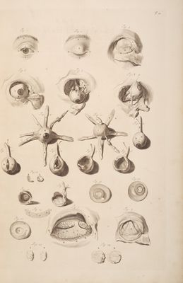
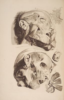
![Fig. f. open’d. * } ; Aid bs AB, The Eye-brow: B, the various Difpofition of its Hairs in Nae this Subject. , C, The Great Canthus of the Eye next the Nofe. D, The Leffler Canthus. R “aG@l E, The Upper Eye-lid. F, The Lower —-— ; G, The White of the Eye cover’d with the Tunica Adnata or Conjunttiva. — _ H, One of the Lachrymal Glands plac’d in the great Canthus of the Eye, call'd Carancula Lachrymalis, and Glandula Lachrymalis Inferior. . Fig. 2. The Eye-lids fhut. _ i A, The Eye-brow, as in the former Figure. C, The Great Canthus of the Eye towards the Nofe. D, The Leffler Canthus. KF, The Superior Palpebra. EF , The Inferior Palpebra. Fig. .2. AA, The Skin with the Mufculus Orbicularis Palpebrarum remov'd. B, The Bone of the Upper Part of the Orbit of the Eye bared. C, The Great Lachrymal Gland involv’d }with Fat. DD, A faint Appearance of the Excretory Duéts of the Lachrymal Glands , by Ber- yichius, call’d Hygroophthalmicos. EE, Divers little Glands interpos’d between the jaft mention’d Dutts. is, . Fig. 4. Parts of the Mufcles of the Eye-lids. tai ditty _ A,Partofthe Mu/culus Aperiens Palpebram Reétus, atits Implantation to the Upper Eye-lid: The Origin of this Mufcle is fharp and flethy at the profoundeft Part of the Orbit , near the Egrefs of the Optick Nerve, accompanying the Reétus Oculi Attollens in its Progrefs , becoming broad, thinand tendonous, as it paffeth over the Superiour Part of the Bulb of the Eye , to its Implantation at the whole Superior Part of the Upper Eye-lid. BC, A Portion of the Upper Part of the Orbicularis Palpebrarum turn’d down, it fill remaining to the Upper Eye-lid: A Defctiption of which Mutfcle will be iriferted in the following Table, Fig. 4. Fe, | nt aee 1 | if 2S. ens t The Lachrymal Glandules, &c. within the Orbit of the Eye, reprefented much bigger than, the Wife, * 5 | AA, The Upper Part of the Bones of the Ortit. BBCC, The Superior Lachrymal Gland, _ DDD, The Vafa Lachrymalia, or Duétus Hygrosphthalmici, whofe Orifices open into the Internal Part of the Palpebre, whence the feparated Liquor, convey’d by the Tubes, iffues to moiften the Palpebre, and External Part of the Bulb of the Eye. EEE, Divers Lachrymal Glands interfpers’d between the laft mention’d Duéts. FF GG, The Cartilages ofthe Cilia joyn’d together with divers Membranes G. Hs The Hairs of the Eye-lids turn’d Upwards, whofe Ramifications appear. 1, Part of the Superior Lachrymal Gland, by Bidloo call’d, Glans Lachrymalis. KK, The Bones of the Nofe broken off, fo that the following Dutts may appeat. _ L, The Dutts, which convey the fuperfluous Moifture of the Va [a Lachrymalia from between the Palpebre; and Bulb of the Eye, into the Foramina Narium: The Orifices of thefe Ducts, appear in the two Papille of the Upper and T_ower Eyc-lid, at the Great Can- thus of the Eye. Fig. t. H. and are evident to the naked Eye , efpecially in thofe, who Cry much,and are call’d, Puncta Lachrymalia: Soon after thefe Two Diséts leave the Great Can- thus of the Eye,they are united into OneTrunk, call’d the Lachrymal.Duét, which defcends in a Foramen of the Second Bone of the Upper Jaw, Tab. 92. Fig.1-D. into the Cavity of the Noftrils. Inthe great Canthus of the Eye arife thofe Tumours ; call’d Egilopes, whofe contain’d Matter , when it degeticrates into an Abfcefs , frequently frames Fiftula’s inthat Part ;and when the Membrane , which compofes the Lachrymal Du@, within the Cavity ofthe Bone , becomes Tumified ; the Paflage of that Duétis rendred impervious ; and Patt of the Humor imploy’d in moiftning the Eye-lids , is hindred in its Difcharge that Way; whence the Cheeks become inflam’d by its running down onthem; in which latter Cafe belides opening the Tumor largely , we muft alfo Perforate the Second Bone of the Upper Jaw or Os Lachrymale, into the Cavity of the Nofe, whereby the Lachrymal Humor will afterwards be difcharg’d: This Operation is beft Praétis'd with a pointed Aétual Cautery fitted with a proper Cannula or Director. The Incifion in thefe Cafes may be made accor. ding to the Direction of the Fibres of the Mu/culus Orbicularis Palpebrarum ; and in ufing the Actual Cautery, great care muft be taken to defend the Palpebre; which we have fometimes feen expos’d to the heated Cannula thro’ the Strugling of the Patient : After this Operation is thus perform’d, it is not neceflary you fhould keep the External Wound open to expeét an Exfoliation of the Fragments of the Bone thro’ it ; but after Three or Four days, when the Callofity isremov’d , andthe Matter difcharg’d , you ought to leffen your Dozils , or Tents, and permit the Sirus to fill with Fleth , and haften a Cica- trice, and the Edges of the perforated Bone will pafs off by the Noftril, as well as that Part of the Bone thruftin by the Cautery. Nor will any great Inconveniency follow if the Healed up Part fhould Impofthumate again, thro’ the moving of the Fragments of the Bone towards the External Wound, if it is again open’d by Incifion to difcharge them ; but fhould you keep the External Wound open long, either by hard Tents, or Efcharo- ticks , you will not only procure a Difcharge of the fuperfluous Tears, or Moifture that way; but the Perforation made in the Os Lachryyale will fill up, and you mutt be oblig’d to repeat the Ufe of the Actual Cautery, or thrufta Probe thro’ it. Ihad almoft forgot to tell you » that after the Firft Incifion made in the External Parts, itis neceflary you fhould pafs your Knife down to the very Bone, and divide the Trunk of a large Artery , and Vein, which pafs that way with the Lachrymal Dug, leaft the Flux of Blood at the time of the Operation fhould fo cool the Cautery , as to preventits Action, | Fig. 6. The Bulb of the Eye lying within the Orbit after the Superior Palpebra is remov’d. AB, The Tunica Adnata plac’d on the Forepart of the Sclerotis. C, The Iris, in whofe Center is the papilla. DD, The Lower Eye-lid, in fitw, together with i Fede ae rene fi gether with Part of the Upper, diffeéted. ¥, The Margin of the Lower Eye-lid, where the Hairs grow out. | Fig. 7 and. &. _ The Mutcles of the Eye, as they ar pear within the Orbit, when clear’d of the Fat and adjacent Parts. ‘ A, The Mufculas Attollens; B, Wig.8.) Mufculns Adducens. C, Deprimens. D, Abducens. . : E , es oe tes of the Bones of the Orbit. , The Tendon of the Mu/culus 08]; ( stoi Infertion behindthe Mu/culus iene Dees Seer ee ce eate eons 1, The External Part of the Bones of the Orbit next the Nofe. sie - : faa or little item ,»on which the Tendon of the Oblique Superior X, Fig. 7. The Qptick Nerve. T! s of th rhe ‘he Fore-parts of the Mufcles of the Right Eye, w | bi clear'd from the Fat , Membranes , and Glands > ae and ee eng enie plane A, Astollens: ; B, Leprimens. C, Adducens, which Bidloo calls Abducens. D, Abducens, which he in like manner miftakes, and calls Adducens, E, Trochlearis Mufculus, or Obliquus Superior cum Trochlea. - ¥, The Trochlea Cartilage , expreft iz itu. Fig. 7. K G, The Mufculus Obliquus Inferior. HHH, The Tunica Adnata,together with another Membranous Tegument detiyg from the Tendons of the Four ftraight Mutcles , mention’d by Realdus Columbus, Lip I, Is fcarce feen , but is plac’din the Center of the Bulb, and diftinguifhes the Pupill , K, Part of the Optick Nerve. t 3 ee Fig: 10.. | : The Back Parts of the Mufcles of the fame Eye, when taken out of the Ozbit oe A, The Mufculus Abducens, or Indignatorius. meee B, Adducens, or Bibitorius. C, Obliquns Inferior, or Breviffimus Oculi Mufculus. D, Attollens, or Superbus. E, Deprimens, or Humilis. F, Obliquus Superior, feu Longiffimus Oculi Mujculus. G, The Trochlea Cartilage. H, A Portion of the Optick Nerve. I, The Back Part of the Bulb of the Eye, compos’d by the Tunica Sclerotis, Fig. 11. The Bulb of the Eye and Optick Nerve free’d from the Mutcles and their Commoy Membranes, fo that the proper Membranes of their Surface appear, A, Part of the Tunica Adnata, which is continued to the Internal Part of the Pah pebre, which can by no means prevent the Retraction of the Eye, when any of the {traight Mufcles Aét, as fome Anatomifts conjecture. BD, The Tunica Sclerotis, C, The Tunica Correa, circumfcrib’d by the Jris, in whofe Center is the Pupilla E, The Optick Nerve cover’d with a Tunick deriv’d from the Dura Mater, j Fig. 12. AA, The Sclerotis open’d, to fhew the Choroeide Tunick immediately under it, B, The Tunica Choroeides. C, The Cornea, Iris, &c. as in the preceding Figure. «Figs 12. Part of the Ligamentum Ciliare view’d with a Microfcope, AABB, The Ligamentum Ciliare confifting of Two Sorts of Fibres; the one extended thro’ its whole Breadth , AA; the other end in the Mid-way BB: Between thefe are plac’d divers Lympheduéts according to Bidloo.. This Mufculous Contexture of the Ligamentum Ciliare moves the Uvea, or Fore-part of the Tuzica Retina compofing the Iris , by which means the Inner Edge of the Jris approaches towards the Center of the Pupilla, or is re. tracted , whereby the Pupil is enlarg’d , or diminith’d according to the different Radiation of Light. In fome Animals, as Cats, exc. we find a Mufculous Struéture inthe Jris alfo, fora more effectual narrowing their Pupils; which Contrivance in thofe Creatures, per- haps , is the more requifite in regard their Horny Tunicks have a Surface not fo prominent in Proportion to the Bulbs of their Eyes, as thofe of other Animals. Fig. 14. The Bulb ofthe Eye together with 4 Portion of the Optick Nerve, where a Divifion of the Tunica Sclerotis together with the Chorotides is made, to exhibit the Tunica Retina, A, The Tunica Retina. Fig. 15. Part of the Optick Nerve together with the Tunicks of the Eye ,after the Humors ( Fig. 19, 20.) are taken out. A, The Inner Surface of the Tunica Retina. Fig. 16. Another View of the Internal and External Surface of the Tunicks of the Eye, after the Humots are difcharg’d. : AA, The Tunica Sclerosis. B, The Cornea. C, Patt of the Optick Nerve. The Internal and F rene e Internal and Fore-part of the Tunicks of the Eve. w t dit charg’d by a tranfverfe Section thro’ the Bulb. 7S) wine Sa A, The Tunica Cornea. C, The Inner Surface of the Jris, next the Ligamentum Ciliare. D, The Tunica Retina Chorocides,and Sclerotis together. Fig. 18. The Inner Surface of the Back Part of the laft mention’d Tunicks of the Eye. A, Part of the Optick Nerve cut off; in which Divifion its Blood-Veffels are expreft, B , The Tunica Sclerotis. | C, The Tunica Retina, in fitu. ee cn Fig. 19, and 20. e Vitreous and Cryitalline Humors of the Eye, when ¢ icks. A, ‘The Cryftalline Humor. | ye; een out of the Tunicks B, The Veftigia of the Ciliar Ligaments on the Vitreous and Edge of the Cryftalline Humor. a C, The Vitreous Humor. Fig. 24. A B, The Cryftalline Humor taken out; A, its Fore-part next the Aqueous Humor; B, as it appears Laterally. The Aqueous Humor cannot eafily be expreft after the Life » Wherefore we fhall here {peak of its Interftice , where it is lodg’d, whereby its Figure is circum{crib’d; its Fore- part is Convext by means of a Concave fram’d by the Cornea in the Center. and Iris it the Circumference; the Back Part of the Aqueous Humor is Concave ,to receive the Co vex Surface of the Cryftalline Humor; it’s fides are Circular, conformable tothe Cavity of the Bulb; whence it appears the Aqueous Humor is Circular in its Circumference, Convext forewards, and Concave backwards, like the following Figure. FA | Ligh 22, A, The External and Fore-part of the Vitreous Humor. B,A Concave in the Vitreous Humor, which receives the Cryftalline Humor. The Tunicle, which is faid to inclofe the Vitreous Humor, do's not appear , but when itis expos’d to the Air; wherefore Dr. Brigzs fuppofes it to be meerly adventitious. Fig. 22. AB » The'Cryftalline Humor dri’d; Bes hy according to fome Anatomitts , calls Tunica Aranca, or Cryftaloeides. | Fie Bis AB, The Vitreous Humor dri’d in like manner; leaving its fuppofed invetting Membrane only. I fhould in this place (as I have hitherto , and fhall hereafter in defcribing of Parts, to which any confiderable Operation of Surgery do’s belong ) {peak of the Couching of Cataracts; but I am afraid I have already tran{greff’d the limits of my Page ; wherefore ! fhall only tell you thatin Prattifing that Operation , the Puncture thro’ the Adzata, ought to be at agreater Diftance from the Pupilla, than Authors commonly direct; and that 4 round Needle is to be preferr’d ; for the edges of the Needle elfe are lyable to wound the Blood-Veflels of the Choroeid Funick largely, and an Extravafation of Blood happens between that Tunick ,and the Sclerosis ; which may be of ill confequence to the Patient.](https://iiif.wellcomecollection.org/image/b33544335_0056.jp2/full/800%2C/0/default.jpg)
 No text description is available for this image
 No text description is available for this image
 No text description is available for this image