A text-book on surgery : general, operative, and mechanical / by John A. Wyeth.
- John Allan Wyeth
- Date:
- 1891
Licence: Public Domain Mark
Credit: A text-book on surgery : general, operative, and mechanical / by John A. Wyeth. Source: Wellcome Collection.
Provider: This material has been provided by the Francis A. Countway Library of Medicine, through the Medical Heritage Library. The original may be consulted at the Francis A. Countway Library of Medicine, Harvard Medical School.
157/912 page 139
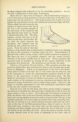
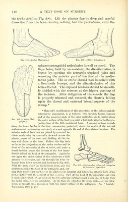
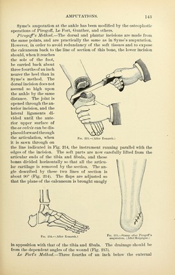
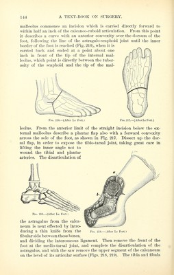
![the flaps trimmed and adjusted as in the preceding operation. Section of tendo A chillis may be done later, if necessary. Medio-Tarsal—Operation of CJiopart.—The dorsal incision is begun on a level with and an inch posterior to the tip of the base of the fifth meta- tarsal bone (for the adult foot). This point is about one fourth of an inch behind the articulation between the cuboid and calcaneum (Figs. 199, 203). With a slight forward convexity the incision is carried across the top of ^ > i \ | the foot to the posterior margin of I ' '■ \ \ the tuberosity of the scaphoid, and ' ; ] \^^^ then directly back from one fourth , - '- T'^- to half an inch (Fig. 198). The skin, ^ / ■; ' // --^/K tendons, vessels, and nerves are di- *'■- ix ^~--'^'-'^'~'~-~^^^'>-^ vided on this line, and the flap lift- ^ \ -. /••''^■^^v/;x- '■--? ■■^ <~^^^i^. ed until the joints between the '^...^^^...^^^^^^■■-■-^►^^ astragalus and scaphoid and the ^^^^^^^^^^..S-^ calcaneum and cuboid are well ex- Fig. 203. posed. From the ends of this first incision a long plantar flap is fashioned by cutting forward, as in shaping the flap for the operation of Lisfranc (Figs. 198,199). Disarticulation is effected with a short, strong scalpel, while forcible extension is employed. The flaps are now to be properly trimmed, and the vessels secured. Division of the tendo Achillis may be done later. When required, this operation may be modified by sawing off the anterior half-inch of the astragalus and calcaneum. The incisions are practically the same. Calcaneo-Astragaloid Disarticulation.—This operation was first sug- gested by Lignerolles, first performed by Textor, but brought into promi- nence by Malgaigne. When in an amputation of the foot at the medio- tarsal joint it is discovered that the os calcis must also be removed, and if the astragalus is sound, the subastragaloid operation should be pre- ferred to the amputation of Syme at the tibio-tarsal joint. By this method a shortening of about two inches is prevented, and, although the under surface of the astragalus is uneven, experience has shown that the pressure is safely distributed, and a useful stump results. Moreover, the degree of mobility maintained at the tibio-astragaloid articulation adds to the ease and comfort of locomotion. Seize the foot with the left hand, and with a strong scalpel commence the incision by dividing the skin and tendo Achillis just at the level of the upper surface of the os calcis. From this point the incision is continued along the fibular side of the foot forward, dividing everything down to the bone, and curving slightly downward until, as it passes below the tip of the external malleolus, it is four tenths of an inch below this point (Fig. 204). The line of incision is now carried directly forward until near the tuber- osity at the base of the fifth metatarsal bone, where it curves to the dor- sum of the foot, crossing to the inner side over the anterior edge of the scaphoid, and then straight down and under the foot a half-inch beyond the middle of the sole (Figs. 205, 206). From this point a straight incision is made directly back to the point of beginning at the inner edge of](https://iiif.wellcomecollection.org/image/b21085201_0157.jp2/full/800%2C/0/default.jpg)
 No text description is available for this image
 No text description is available for this image
 No text description is available for this image