On mycetoma, or the fungus disease of India / by H. Vandyke Carter.
- Henry Vandyke Carter
- Date:
- 1874
Licence: Public Domain Mark
Credit: On mycetoma, or the fungus disease of India / by H. Vandyke Carter. Source: Wellcome Collection.
Provider: This material has been provided by The Royal College of Surgeons of England. The original may be consulted at The Royal College of Surgeons of England.
132/172
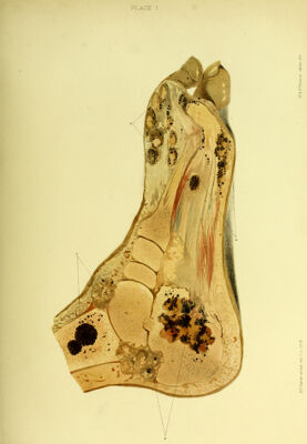
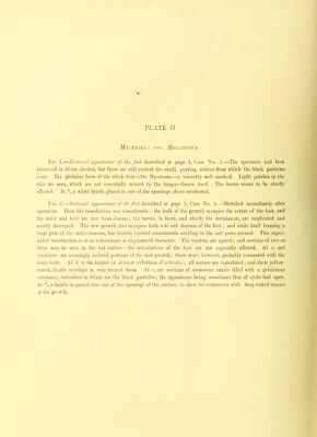
![A PLATE II Mycetoma ; var. Melanotica Tig. 1.—External appearance of the foot described at page 4, Case jVo. 2.—The s])cciinen had been ])reserved in dilute alcoliol, but there are still evident the small, pouting orifices from which the black particles issue. The globular form of the whole foot—the Mycetoma—is tolerably well marked. Light patches in the skin are seen, which are not essentially related to the fungus-disease itself. The tarsus seems to be chiefly affected. At *, a white bristle placed in one of the openings above mentioned. Tig. 2.—Sectional appearance of the foot described at page .5, Case No. !•.—Sketched immediately after operation. Here the tumefaction was considerable : the bulk of the growth occupies the centre of the foot, and the ankle and heel are free from disease; the tarsus, in front, and chiefly the metatarsus, are implicated and mostly destroyed. The new growth also occupies both sole and dorsum of the foot; and while itself forming a large part of the entire tumour, has besides excited considerable swelling in the soft parts around. This super- added tumefaction is of an a-dematous or elephantoid character. The tendons are spared; and sections of two or three may be seen in the cut surface; the articulations of the foot are not especially affected. At a, and elsewhere, are seemingly isolated portions of the new growth; these were, however, probably connected with the main body. At i, is the largest or densest collection of sclerotia ; all masses are capsulated ; and their yellow- tinted, friable envelope is seen around them. At c, are sections of numerous canals filled with a gelatinous substance, imbedded in which are the black particles; the appearance being sometimes that of cysts laid open. At a bristle is passed into one of the openings of the surface, to show its connection with deep-seated masses of the growth.](https://iiif.wellcomecollection.org/image/b22372635_0134.jp2/full/800%2C/0/default.jpg)