On mycetoma, or the fungus disease of India / by H. Vandyke Carter.
- Henry Vandyke Carter
- Date:
- 1874
Licence: Public Domain Mark
Credit: On mycetoma, or the fungus disease of India / by H. Vandyke Carter. Source: Wellcome Collection.
Provider: This material has been provided by The Royal College of Surgeons of England. The original may be consulted at The Royal College of Surgeons of England.
79/172 (page 69)
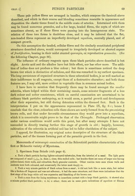
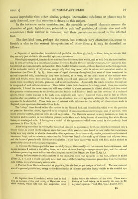
![These pale yellow fibres are arranged in bundles, which compose the fasciculi above described, and which in their course and blending sometimes resemble in appearance and disposition the elastic tissue found in the middle coats of arteries. Intermixed with them may be found numerous granules, and a few large, beaded fibres, the septa of which are sometimes absent, as if these fibres were passing into the homogeneous state. The relation of these two forms is doubtless close, and it may be inferred that the flat, non-beaded fibres represent an imperfectly developed mycelium, centrally placed, and of compact growth. On this assumption the beaded, cellular fibres and the similarly constituted peripheral structures described above, would correspond to irregularly developed or aborted organs of fructification, bearing in their midst stunted sporangia, or reproductive gemmules. {See the following Ch«,pter, page 86). The influence of ordinary reagents upon these black particles above described is but slight. Acetic acid and the alkalies have but little effect, nor has ether more. The addi- tion of iodine does not produce a blue colour ; but there is sometimes noticed the deep brown tint which is distinctive of cellulose, and this was remarked in my earliest specimen. The long persistence of organized structure in these sclerotioid bodies, is as well marked as their indifference to all reagents, except those of a destructive character; and both these features are, I may add, more evident in vegetable than in common animal bodies. I have here to mention that frequently there may be found amongst the smaller sclerotia, where lodged within their containing canals, some minuter fragments of a less dark colour and softer consistence, which on careful examination are ascertained to be ordinary black particles undergoing, as it would seem, a partial growth and development after their separation, but still during detention within the diseased foot. Such is the interpretation I put on the appearances represented in Plate IX, fig. 6 c; hence I conclude that the clear, colourless cells there shown are newly formed from the pre-existing brown cells, and that under favorable circumstances they would grow into a mycelium, which it is conceivable might prove to be that of the Chionyphe. Prolonged observation under various conditions would settle this point, but after many attempts I have not succeeded in directly tracing further this stage of growth in situ; happily, however, cultivation of the sclerotia in artificial soil has led to fuller elucidation of the subject. I append, for illustration, my original notes descriptive of the structure of the black particles, and of the tissues forming part of the parasitic growth in the foot. Memoranda of microscopic examination of the Sclerotioid particles characteristic of the first or Melanotic variety of Mycetoma. 1. Specimen from Scinde {vide page 4). a. Took a black particle having lighter-coloured tuhefcules, from the interior] of a canal. The light parts are composed of small in. diam.), clear, thin-walled cells; hut besides there are some of larger size having yellow-tinted, thick walls, and colourless, finely granular contents. Other vesicles were seen whose walls and contents were both coloured, and occasionally the latter most deeply so. h. Took a very minute particle of black colour from a canal; it did not look like a mere stain, hut rather like a broken-off fragment and was not adherent; it had the same structure, and there were indications that the first stage of the large white cell was expansion and blanching of the brown one. c. Took a stain from the lining membrane in immediate contact with a large black particle; it showed also a minute perfect black particle; + Ac + ether; there are in some parts indications of mycelium or fibrous](https://iiif.wellcomecollection.org/image/b22372635_0081.jp2/full/800%2C/0/default.jpg)