The elements of embryology. / By M. Foster ... and Francis M. Balfour.
- Michael Foster
- Date:
- 1874
Licence: Public Domain Mark
Credit: The elements of embryology. / By M. Foster ... and Francis M. Balfour. Source: Wellcome Collection.
Provider: This material has been provided by The University of Leeds Library. The original may be consulted at The University of Leeds Library.
126/336 (page 100)
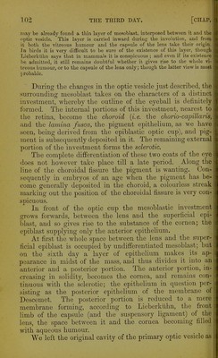
![order to be seen must be looked for on the under surface of the optic vesicle. In this position it is readily recog- nized in the transparent embryo of the third day, Figs. 25' and 29. Bearing in mind these relations of the gap to the optic stalk, the reader will understand how sections of the optic vesicle at this stage present very different appearances according to the plane in which the sections are taken. When the head of the chick is viewed from underneath as a transparent object the eye presents very much the ap- pearance represented in the diagram Fig, 29. D D. Diagrammatic section taken perpendicular to the plane of the paper, along the line y, y. Fig. 29. The stalk is not seen, the section falling quite out of its region. rA, hollow of optic cup filled with vitreous humour; other letters as in Fig. 2] B. E. Section taken parallel to the plane of paper through Fig. 29, so far behind the front surface of the eye as to shave off a small portion of the posterior surface of the lens I, but so far in front as not to be can-led at all through the stalk. Letters as before; /, the choroidal fissure. F. Section along the line z, 2, perpendicular to the plane of the paper, to shew the choroidal fissure /, and the continuity of the cavity of the optic stalk with that of the primary optic vesicle. Had this section been taken a little to one side of the line z, s, the wall of the optic cup would have extended up to the lens below as well as above. Letters as before. A section of such an eye taken along the line y, per- pendicular to the plane of the paper, would give a figure corresponding to that of Fig. 30 D. The lens, the cavity and double walls of the secondary vesicle, the remains of the primary cavity, would all be represented (the superficial epiblast of the head would also be shewn); but there would be nothing seen of either the stalk or the fissure. If on the other hand the section were taken in a plane parallel to the plane of the paper, at some distance above the](https://iiif.wellcomecollection.org/image/b2150684x_0128.jp2/full/800%2C/0/default.jpg)