The elements of embryology. / By M. Foster ... and Francis M. Balfour.
- Michael Foster
- Date:
- 1874
Licence: Public Domain Mark
Credit: The elements of embryology. / By M. Foster ... and Francis M. Balfour. Source: Wellcome Collection.
Provider: This material has been provided by The University of Leeds Library. The original may be consulted at The University of Leeds Library.
175/336 (page 149)
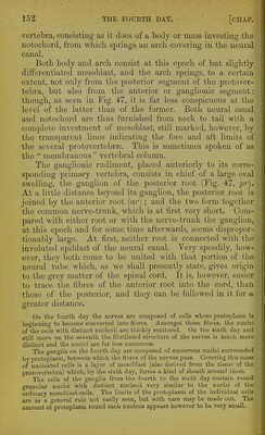
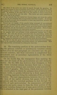
![the wall of the cloaca: with this they unite, and form together a solid spherical body, bearing on its external surface a median furrow, indicating its double origin. A narrow diverticulum of hypoblast now passes into the mass and forms within ic a cavity, which is at first small and, corresponding to the ex- ternal contour of the body, to a certain extent double. The hypoblast diverticulum grows rapidly, while its mesoblastic covering remains nearly stationary, so that the mesoblast finally comes to form a thin coating only over the hypoblast. His [op. cit.) gives a somewhat elaborate and complicated account of the development of the allantois; which is accepted by Waldeyer {Eierstoclc unci .Ei) and Bornhaupt (Untersuchung uber die Entwickelung des Urino-genitalsystems helm Hiihnchen, Eiga, 1867). It appears to be nearly the same as the fuller account given by Dobrynin {Ueber die erste Anlage der Allantois. Sitz. der k. Akad. Wien, Bd. 64, 1871), of which the following is an abstract. When the first commencement of the hind fold takes place, immediately beyond the point where the hypoblast turns back to assume its normal direction over the yolk-sac, a narrow diverticulum which points backwards and some- what upwards is formed by a special flexure of the splanchnopleure. The open end of the diverticulum. Fig. 49, AIL, looks forwards towards the wide opening connecting the digestive tract with the yolk-sac; its blind end points directly towards the pleuroperitoneal cavity. This diverticulum is the commencing allantois. It is lined by hypoblast, while its exterior is composed of the mesoblast of the splanchnopleure. As the folding in to form the digestive tract increases, the diverticulum alters riG. 50* ] Longitudinal Section op the Tail-end op an Embexo Chick at the MIDDLE OF THE Thikd Day (Dobrynin). tt. the tail; the line of reference points to the axial mesoblast at the tail, x'. epiblast. SO. somatopleure. m. mesoblast to form the body wall, V. commencing amniotic fold. JIp. space between the true and the false amnion, pp. Pleuroperitoneal cavity. Spl. splanchnopleure. 6. Cloacal enlargement of the alimentary canal. Dd. dorsal wall of the alimentary canal. All. vesicle of the allantois having a wide opening into the alimen- tary canal.](https://iiif.wellcomecollection.org/image/b2150684x_0179.jp2/full/800%2C/0/default.jpg)