Histology of the cholera evacuations in man and the lower animals / by W. Lauder Lindsay, M.D.
- William Lauder Lindsay
- Date:
- 1856
Licence: Public Domain Mark
Credit: Histology of the cholera evacuations in man and the lower animals / by W. Lauder Lindsay, M.D. Source: Wellcome Collection.
Provider: This material has been provided by The University of Glasgow Library. The original may be consulted at The University of Glasgow Library.
8/40 page 8
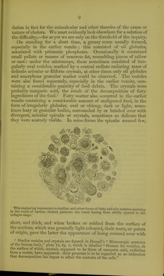
![lished, as delineative of the bodies in question, by tlieir original discoverers. I believe that potatoes, oatmeal, bread, and the veget- ables of common broth, will furnish most of the forms of the once famed annular bodies ; that they are not, therefore, fungoid in their nature or origin; and that they have no essential or causative relation to cholera. I have found them equally in other diseases— as in the stools of diarrhoea and dysentery. It is unnecessary to specify, in detail, the forms of these cellular bodies, observed by myself.^ Many of them are simple globular cells, containing chlorophylle granules, aggregated regularly or irregularly round a central nucleus. This is the character of the gonidia of lichens— the green coloured cells lying immediately below the cortical layer of the thallus: hence, in former publications, in order to economise space and words, and to indicate their general appearance, I deno- minated these bodies gonidic. When- emptied of their contents, they are delicate hyaline vesicles, and appear often as mere circles or rings, enclosing a free central area. Sometimes there is an inner dotted ring, probably produced by a puckering of the cell wall. This cell wall sometimes disappears, and the chlorophylle grains may then be found aggregated circularly round their nucleus, in regular masses, or free and intermixed with granular debris. Others of these bodies are of larger size, but very irregular shape : their walls are thick, and variously coloured, especially brown. Starch glo- bules, partially broken up, are probably a common form of annular bodies : in this condition they frequently resemble the shrivelled sporangia of ferns. The non-action of iodine is not, I think, a sufficient disproof of such bodies being of an amyloid nature; for I have already mentioned that its usual reaction is sometimes absent, where the corpuscles otherwise bear indubitable marks of being starch. I have never seen annular bodies produced from s])iral or annular vessels of plants, as was suggested in the report of the College of Physicians, London, in reference to the bodies described and figured by Brittan and Swayne of Bristol, though such vessels themselves are far from uncommon. Nor have I been able to trace any of them to medicines, for they occurred equally in cases where no medicines were given, or prior to their administra- tion. Moreover, they were comparatively seldom found in the collapse stools, occurring chiefly in those of the reaction and fever stages; and their presence was coincident with the appearance of less equivocal forms of food debris. It will be evident, then, that I can see no satisfactory groundwork for the fungus theory of cholera, which I am not a little surprised to find still possesses powerful advocates.^ We shall, hereafter, see that there is equally little foun- 1 Drawings and descriptions will be found in the Association Med. Journal, April 14, 1854. „ , , Fide Professor Daubeny of Oxford, On the influence of the lower veget- able organisms in the production of epidemic diseases.—iS^cZm. New Philosoph. Joitrtial, July 1855,](https://iiif.wellcomecollection.org/image/b21477875_0008.jp2/full/800%2C/0/default.jpg)