Auricular fibrillation and its relationship to clinical irregularity of the heart / by Thomas Lewis.
- Thomas Lewis
- Date:
- [1910?]
Licence: In copyright
Credit: Auricular fibrillation and its relationship to clinical irregularity of the heart / by Thomas Lewis. Source: Wellcome Collection.
61/80 page 363
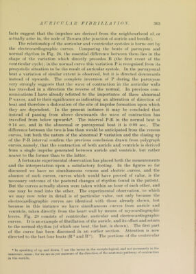
![facts suggest that the impulses are derived from the luughhourliood of, or actually arise iu, the node of Tawara (the junction of auricle and hiindle). The relationship of the auricular and ventricidar systoles is borne out h}' the electrocardiogra])hic curves. Comparing the beats of ])arox3'sm and normal rhythm in Fig. 2(), the essential difference between them lies in the shape of the variation which directly ])recedes H (the first event of the ventricular cycle); in the normal curve this variation P is recognised from its presystolic situation to he the result of auricular systole. In the ])aroxysmal heat a variation of similar extent is observed, hut it is directed downwards instead of upwards. The com])lete inversion of P during the ])aroxysm very strongly suggests that the wave of contraction in the auricular walls has travelled in a direction the reverse of the normal. In ])revious com- munications I have already referred to the importance of these abnormal P w aves, and to their significance as indicating an alteration of direction of beat and therefore a dislocation of the site of impulse formation upon which they are dependent. In the present instance it may be concluded that instead of passing from above downwards the wave of contraction has travelled from below upwards*. The interval P-R in the normal beat is 0-14 sec. and in the abnormal or paroxysmal beat it is 0-08 sec.. The difference between the two is less than would be anticipated from the venous curves, but both the nature of the abnormal P variation and the closing up of the P-R interval confirm the previous conclusion drawn from ])olygraph curves, namely, that the contraction of both auricle and, ventricle is derived from a single impulse generated between auricle and ventricle, but rather nearer to the former than to the latter. A fortunate experimental observation has placed both the measurements and the interpretation upon a satisfactory footing. In the figures so far discussed we have no simultaneous venous and electric curves, and the absence of such curves, curves which would have ])roved of value, is the necessary outcome of the postural changes of rhythm found in the patient. But the curv'cs actually shown were taken within an hour of each other, and one may be read into the other. The experimental observation, to which we may now refer in detail is of ])articular value, not only because the electrocardiogra])hic curves are identical with those already shown, but because in this instance we have simultaneous curves from auricle and ventricle, taken directly from the heart wall by means of myocardiograi)hic levers. Fig. 2!> consists of ventricular, auricular and electrocardiogra])hic curves. It is an exam])le of fibrillation of the auricle, and its offset and return to the normal rhythm (of which one beat, the last, is show n). I he first ])art of the curve has l)een discussed in an earlier section. Attention is now directed to the last four beats (R'^ and R’). The portions of the ventricular * In apeakincr of np and down, 1 uso the terms in the inor|)liological, and not neee.s.sanly in Uie anatomic, sense ; for we are as yet unaware of the direction of the anatomic pathway of contraction in the auricle.](https://iiif.wellcomecollection.org/image/b29000610_0061.jp2/full/800%2C/0/default.jpg)