Auricular fibrillation and its relationship to clinical irregularity of the heart / by Thomas Lewis.
- Thomas Lewis
- Date:
- [1910?]
Licence: In copyright
Credit: Auricular fibrillation and its relationship to clinical irregularity of the heart / by Thomas Lewis. Source: Wellcome Collection.
63/80 page 365
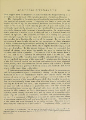
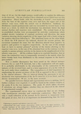
![F AURICULAR FIBRILLATION. 3(35 time of -01 sec. for the single journey would suffice to explain the dilference in the interv^als. On one occasion 1 have obtained curves which bear out this explanation. Direct leads, with the electrodes of Gotch'^, were instituted from the base of the superior vena cava and from the upper end of the inferior vena cava, while the auricle was in fibrillation. Suddenly the fibrillation ceased spontaneously and was succeeded by several beats of the heart as a whole, in which the mechanism was abnormal, before the final normal rhythm and sequence were established. The normal beats of the re-established rhythm were accompanied by auricular contractions which yielded electric variations of constant excursion and direction, the main variation showing electro-negativity of the upper lead with a lesser swing in the positive direction following it. The abnormal beats on the other hand, in which the P-R interval was reduced, showed the reverse picture, the excursions remained the same but the direction altered from negative to positive and positive to negative respectively. In the case of the normal beat we have to assume primary* activity of the tissues abutting on the superior vena cava; in the case of the abnormal beat on the contrary we have equally to assume primary activity in the tissues in the neighbourhood of the inferior vena cava. All the evidence leads us to conclude therefore, that at times the contraction wave is temporarily reversed in the auricle when the latter breaks back from fibrillation to the normal response to the heart’s pace-makerf. Now a similar discrepancy has been noted in the clinical instance between the a-c and P-K intervals, but a like explanation will not apply. It is probably attributable to the abnormality of the contraction in the instance of the reversed beat, and to a later a])pearance of a in the veins of the neck under these conditions. The com])arison is of chief imjjortance in emphasising the greater accuracy of the P-R as o[)poscd to the u-c interval in the clinical instance. The a-c interval during the ])aroxysm is not an absolutely true representation of the As-\s interval, it is too short, and the electric measurement, -08 sec., is the more accurate. The parallel between clinical and experimental curves is striking, but if further evidence were required, it would be found in the relative heights of the })eaks R in the two instances. The ]»eaks R which follow inverted P variations are always higher, whether they are observed in the clinical or experimental example. The experimental curves show us conclusively that we arc dealing with simultaneous auricular and ventricular contraction. That tlicie Is only a small reduction of the normal As-^s interval is immaterial fiom this point of view, but it is of importance in demonstrating that the level * Referring to svn)orior and inferior vena eava only. t \Vinterl)erg«‘ ha.s made certain .)l)s«'rvalions which tend to tlic same auricular inii»ul.ses, at the escape from fil)rillation. are not necessari y “sinus,” they may arise in other portions of the auncidar walls. conclusam. '1 he generated in the](https://iiif.wellcomecollection.org/image/b29000610_0063.jp2/full/800%2C/0/default.jpg)


 No text description is available for this image
 No text description is available for this image
 No text description is available for this image