Atlas of cutaneous morbid histology / by Max Joseph and J. B. Van Deventer.
- Joseph, Max, 1860-1933.
- Date:
- 1906
Licence: In copyright
Credit: Atlas of cutaneous morbid histology / by Max Joseph and J. B. Van Deventer. Source: Wellcome Collection.
Provider: This material has been provided by The University of Glasgow Library. The original may be consulted at The University of Glasgow Library.
30/114 page 8
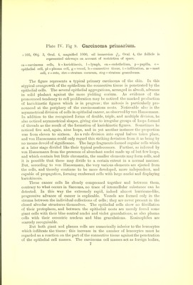
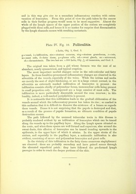
![and in this way give rise to a secondary inflammatory reaction with extra- vasation of leucocytes. From this point of view the path taken by the cancer cells in their further progress would seem to be most suggestive. Almost the whole of the lymph spaces of the upper portion of the corium are completely plugged with these cells, and hence it is no matter for surprise that dissemination by the lymph channels ensues with resulting metastasis. Plate IV. Fig. 10. Folliculitis. X Leitz, Obj. 3, Ocul. 3. g = vessel, i = infiltration, strc = stratum corneum, strg = stratum granulosum, r = rete, m = mast cells, f = fatty tissue, p = plasma cells, sch = sweat glands, mc = micrococci, ch = chromatotaxis. The two last are x 800 Leitz, Obj. oil immersion, and Ocul. 3. The original was taken from a girl whose forearm was the seat of an abundant, nearly symmetrical, and typical eruption. The most important morbid changes occur in the sub-cuticular and fatty layers. In these localities pronounced inflammatory changes are observed in the adventitia of the vessels, especially of the veins. While the intima and media are merely the seat of slight thickening, or are to a large extent normal, in the adventitia an extremely marked infiltration of leucocytes is present: the infiltration consists chiefly of polynuclear forms, mononuclear cells being present in small proportion only. Interspersed a]:e a large number of mast cells. The infiltration is more particularly discernible around the vasa vasorum; in this locality, indeed, a well-marked periphlebitis is present. It is noticeable that this infiltration leads to the gradual obliteration of the vessels around which the inflammatory process has taken its rise ; so marked is this occlusion that it is difiicult to discover the existence of a lumen as regards these vessels. Hence it is not surprising that the papule is destroyed and that, as a result of the process of papule formation, an ultimate condition of papule destruction ensues. The path followed by the assumed tubercular toxin in this disease is probably rendered evident by an infiltration of leucocytes which can be traced along the vessels up to the papillary layer. Along the lymph spaces situated on the route, and when these spaces are well formed and capacious, also around the sweat ducts, this effusion of leucocytes can be traced trending upwards to the epidermis, to the upper layer of which it attains. In the upper strata of the corium, and especially in the papillary layer, a noticeable infiltration is also evident, consisting of mono- and polynuclear leucocytes and giant cells. In the vicinity of this infiltration in the fatty tissue numerous staphylococci are observed : these are probably secondary and have gained access through the ulcerated superficial patch: they have followed the pre-formed lymph passages in order to reach the deeper portions of the skin.](https://iiif.wellcomecollection.org/image/b21463621_0030.jp2/full/800%2C/0/default.jpg)
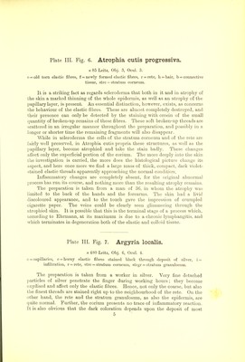

 No text description is available for this image
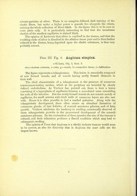
 No text description is available for this image
 No text description is available for this image