A text-book of the diseases of the ear / by Josef Gruber; translated from the second German edition by special permission of the author, and edited by Edward Law and by Coleman Jewell.
- Josef Gruber
- Date:
- 1890
Licence: Public Domain Mark
Credit: A text-book of the diseases of the ear / by Josef Gruber; translated from the second German edition by special permission of the author, and edited by Edward Law and by Coleman Jewell. Source: Wellcome Collection.
Provider: This material has been provided by University of Bristol Library. The original may be consulted at University of Bristol Library.
29/618
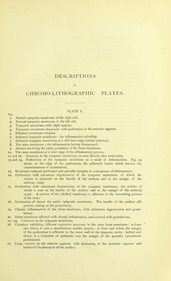
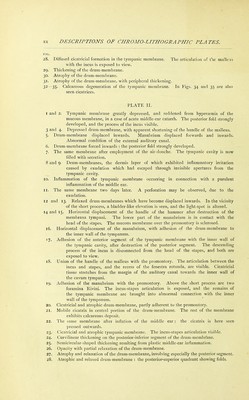
![FIG. 29. The same membrane as in Fig. 28, as seen during inflation by the Valsalvan process. 30. Relaxed tympanic membrane. The relaxed parts are in distinct folds on the posterior segment. 31. The same membrane during Valsalvan inflation : the folds have now disappeared. 32. An atrophic drum-membrane with its posterior segment considerably relaxed. The relaxed portion is displaced backwards, upwards, and inwards; and exhibits folds at the lower part posteriorly. In the pos'erior-superior quadrant mav be perceived the incus-stapes articulation and the chorda tympani. 33. The same membrane as observed during Valsalvan inflation. 34. Atrophic and relaxed tympanic membrane. On the posterior segment is seen a curved line, representing the place of attachment of a pseudo-membrane, by which an abnormal connection exists between the membrana tympani and the inner wall of the tympanic cavity. The drum-membrane is folded above and below this line. 135. • The same membrane during inflation by the Valsalvan method. The relaxed parts appear bulged out like a bladder, and an oblique furrow is here visible, corre- sponding to the attachment of the above-mentioned false membrane. INDEX OF ILLUSTRATIONS IN THE TEXT. K]G. PAGE 1. Squamous portion of the temporal bone, seen from without ----- 4 2. Squamous portion of the temporal bone, seen from within ----- 4 3. Tympanic ring - - , - . - - 5 4. Anterior surface of the petrous portion of the temporal bone ----- 6 5. Posterior surface of the petrous portion --------- 7 6. Upper surface of the petrous portion --------- 7 7. Under surface of the petrous portion --------- g 8. Section through the mastoid process of an infant ------- 13 9. Temporal bone of an infant, seen from the outer side - - - - 14 10. Temporal bone of an infant, seen from the inner side ------ 14 11. Temporal bone at birth - - - - - - 18 12. Temporal bone from a boy, two and a half years old ... . - 18 13. Temporal bone of an adult. The under surface of the petrous portion is also seen - 19 14. Temporal bone of an adult, seen from the outer side - 20 15. Temporal bone of an adult, viewed from the inner surface - - - - - 21 16. Section through the mastoid portion at birth - ------- 23 17. Section through the mastoid portion of a child, aged two years - - - - 23 18. Section of the mastoid portion of a boy, aged three years 23 19. Section through the mastoid portion of a man, aged thirty - - 24 20. Section through the mastoid portion of a man, aged thirty ----- 24 21. Section through the mastoid portion and tympanic cavity of a man, aged thirty - 24 22. Section through the mastoid portion of a man, aged thirty 24 23. Section through an entirely diploe'tic mastoid process of a man, aged thirty - - 25 24. View of the anterior surface of the petrous bone united with the mastoid - - - 27 25. Outer extremity of the internal auditory canal ------ - 30 26. Bony labyrinth of the right ear - - - - - - - - - - 35 27. Right bony labyrinth with the canals laid open ------- 36 28. Section parallel with the axis of the cochlea -------- 37 29. Pinna - - - 41 30. Section through the ear, parallel with the long axis of the external auditory canal - 43](https://iiif.wellcomecollection.org/image/b21446775_0029.jp2/full/800%2C/0/default.jpg)