Practical anatomy: a manual of dissections / by Christopher Heath.
- Christopher Heath
- Date:
- 1870
Licence: Public Domain Mark
Credit: Practical anatomy: a manual of dissections / by Christopher Heath. Source: Wellcome Collection.
Provider: This material has been provided by the Francis A. Countway Library of Medicine, through the Medical Heritage Library. The original may be consulted at the Francis A. Countway Library of Medicine, Harvard Medical School.
421/600 (page 411)
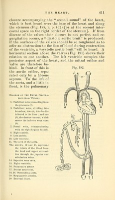
![closure accorapanj'ing the second sound of the heart, which is best heard over the base of the heart and along the sternum (Fig. 188, B, p. 402) [or at the second inter- costal space on the right border of the sternum]. If from disease of the valves their closure is not perfect and re- gurgitation occurs, a diastolic aortic bruit is produced: if the surfaces of the valves should be so roughened as to offer an obstruction to the flow of blood during contraction of the ventricle, a sj^stolic aortic bruit will be heard. A horizontal section above the valves (Fig. 191) shows their relation to one another. The left ventricle occupies the posterior aspect of the heart, and the mitral orifice and valve are therefore be- hind. In front of this is Fig. 192. the aortic orifice, sepa- / /^> rated only by a fibrous ■' septum. To the left of the aorta, and a little in front, is the pulmonary Diagram of the Fcetal Ciroula- TiON (from Wilson). 1. Umbilical vein proceeding from the placenta (2). 3. Umbilical vein, dividing into branches; two (4, 4) to be dis- tributed to the liver ; and one (5), the ductus venosus, which enters the inferior vena cava (6). 7. Portal vein, communicating with the right hepatic branch. 8. Right auricle. 9. Left auricle. 10. Left ventricle. 11. The arch of the aorta. The arrows, 12 and 13, repre-isent the return of the blood from the head and upper exti'emi- ties through the jugular and subclavian veins. 14 Superior vena cava. 1.5. Riglit ventricle. 16. Pulmonary artery 17. Ductus arteriosus. IS, 18. Descending aorta. 19. Hypogastric arteries. 20. External iliacs. \J \>](https://iiif.wellcomecollection.org/image/b21057679_0421.jp2/full/800%2C/0/default.jpg)