Practical anatomy: a manual of dissections / by Christopher Heath.
- Christopher Heath
- Date:
- 1870
Licence: Public Domain Mark
Credit: Practical anatomy: a manual of dissections / by Christopher Heath. Source: Wellcome Collection.
Provider: This material has been provided by the Francis A. Countway Library of Medicine, through the Medical Heritage Library. The original may be consulted at the Francis A. Countway Library of Medicine, Harvard Medical School.
433/600 (page 423)
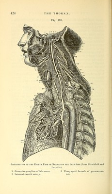
![great variations in size, and when much lij'pertropliied constitnte Bronchocele, or Goitre. The thja-oid body is covered by the sterno-hyoid and thyroid muscles, and occasionally a few muscular fibres pass from the li3oid bone to the isthmus, constituting the levator glandulse thyroidese of Soemmering. It is abun- dantly supplied with blood b}^ the superior thyroid and inferior thj/roid arteries of each side, and occasionally b}'^ an additional branch from the innominate. The arteries freel}^ anastomose in the substance of the bod}^, and return their blood by three veins on each side, A'iz., the superior and middle thyroid which join the in- ternal jugular vein, and the inferior thyroid which has been traced down the front of the trachea to the innominate vein. The thjn'oid body is composed of numerous closed vesi- cles containing a yellow fluid, but its function is not under- stood. [It belongs probably to the lymphatic system.] The right luug is to be drawn forward and the pleura divided where it is reflected from the lung to the wall of the thorax, and the parts in the posterior mediastinum are to be cleaned. The muscular oesophagus will be at once seen, and the right pneumogastric nerve is to be traced to it and to the back of the right bronchus. On displacing the oesophagus the side of the thoracic aorta will come into view, but it will be better seen in the dissection of the left side. The vena azygos major will be seen to the right of the aorta, and between the two will be found the slender and collapsed thoracic duct. The intercostal vessels will be seen crossing the back of the space, and near the diaphragm will be found the splanchnic nerves from the sympathetic cord, which is itself outside the mediastinum. The Posterior Mediastinum (Fig. 185, p. 398) is the interpleural space behind the pericardium, bounded by the vertebrae behind, the pericardium in front, and the reflec- tion of the pleura on each side. It contains the trachea, oesophagus, and the two pneumogastric nerves ; the thoracic aorta, vena azygos major and thoracic duct; and, at the lower part, the great splanchic nerves. The (Esophagus (Fig. 194, 29, p. 420) is a muscular tube continuous with the pharynx. It begins opposite to the 5th cervical vertebra, and can now be seen to lie to the left side of the median line in the anterior triangle. It then passes through the superior aperture of the thorax, being in relation with the left common carotid artery, and reaches](https://iiif.wellcomecollection.org/image/b21057679_0433.jp2/full/800%2C/0/default.jpg)