Practical anatomy: a manual of dissections / by Christopher Heath.
- Christopher Heath
- Date:
- 1870
Licence: Public Domain Mark
Credit: Practical anatomy: a manual of dissections / by Christopher Heath. Source: Wellcome Collection.
Provider: This material has been provided by the Francis A. Countway Library of Medicine, through the Medical Heritage Library. The original may be consulted at the Francis A. Countway Library of Medicine, Harvard Medical School.
439/600 (page 429)
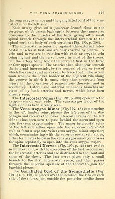
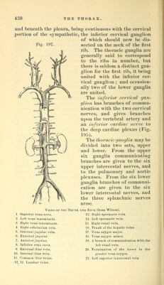
![the vena azygos minor and the gangliated cord of the sym- pathetic on the left side. Each arter}^ gives off a jjosfeinor branch close to the vertebra?, which passes backwards between the transverse processes to the mnscles of the back, giving off a small spinal branch through the intervertebral foramen to the spinal cord and bod}^ of each vertebra (Fig. 185, p. 398). The intercostal arteries lie against the external inter- costal muscles at first, and are only covered by pleura. A vein and nerve are in relation with each artery, the vein being highest and the nerve lowest in most of the spaces, but the artery being below the nerve at first in the three or four upper spaces. The arteries then disappear beneath the internal intercostals, by the removal of one or two of Avhich the vessels and nerves can be traced out. The artery soon reaches the lower border of the adjacent rib, along the groove in which it runs, being thus protected from injnrj' in the operation of paracentesis thoracis [and in accidents]. Lateral and anterior cutaneous branches ai'e given off by both arteries and nerves, which have been already seen. The Intercostal Veins (Fig. 19t, p. 430) open into the azygos vein on each side. The vena azygos major of the right side has been already seen. The Vena Azygos Minor (Fig. 197, i8) commencing in the left lumbar veins, pierces the left crus of the dia- phragm and receives the lower intercostal veins of the left side; it has been seen to pass behind the aorta and open into the vena azygos major. The upper intercostal veins of the left side either open into the sui^erior intercostal vein or form a separate vein (vena azj^gos minor superior) which, communicating with the superior costal vein above, either terminates below in the vena azygos minor, or crosses the spine separatel}^ to open into the vena azygos major. The Intercostal Nerves (Fig. 195, p. 424) are twelve in number, and, with the exception of the first, accompany the intercostal arteries and are distributed to the front and sides of the chest. The first nerve gives only a small branch to the first intercostal space, and then passes through the superior aperture of the thorax to join the brachial plexus. The Gangliated Cord of the Sympathetic (Fig. 194, 31, p. 420) is placed over the heads of the ribs on each side of the thorax just outside the posterior mediastinum](https://iiif.wellcomecollection.org/image/b21057679_0439.jp2/full/800%2C/0/default.jpg)